


Fibrous joints
Fibrous joints are of three types: suture, gomphosis and syndesmosis.
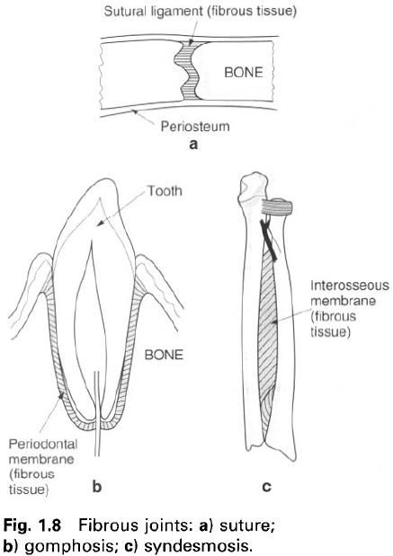
Suture(figure
a)
This is a form of fibrous joint that exists
between the bones of the skull. They permit no movement as the edges of the
articulating bones are often highly serrated, as well as being united by an
intermediate layer of fibrous tissue. Either side of this fibrous tissue the
inner and outer periosteal layers of the bones are continuous, and in fact
constitute the main bond between them.
The sutures are not permanent joints, as they
usually become partially obliterated when age increases beyond 30 years.
Gomphosis(figure
b)
In this form of fibrous joint a peg fits into a
socket, being held in place by a fibrous ligament or band; the roots of the
teeth being held within their sockets in the maxilla and mandible are such
examples.
Syndesmosis(figure
c)
In a syndesmosis the uniting fibrous tissue is
greater in amount than in a suture, forming a ligament or an interosseous
membrane. Examples in the adult are the inferior tibiofibular joint where the
two bones are joined together by an interosseus ligament, and the interosseus
membrane between the radius and ulna. Flexibility of the membrane or twisting
and stretching of the ligament permit movement at the joint. However, the
movement allowed is restricted and controlled.
Cartilaginous joints
In cartilaginous joints the two bones are
united by a continuous pad of cartilage. There are two types of such joint, primary and secondary cartilaginous(synchondrosis
and symphysis respectively).
Primary
cartilaginous(figure a)
Between the ends of the bone involved in the
joint is a continuous layer of hyaline cartilage. These joints occur at the
epiphyseal growth plates of growing and developing bone, and obviously become
obliterated with fusion of the two parts(diaphysis and epiphysis). Because the
plate of hyaline cartilage is relatively rigid, such joints exhibit no
movement. However there is one such joint in the adult, which is slightly modified
because, by virtue of its structure, it enables slight movement to occur. This
is the first sternocostal joint.
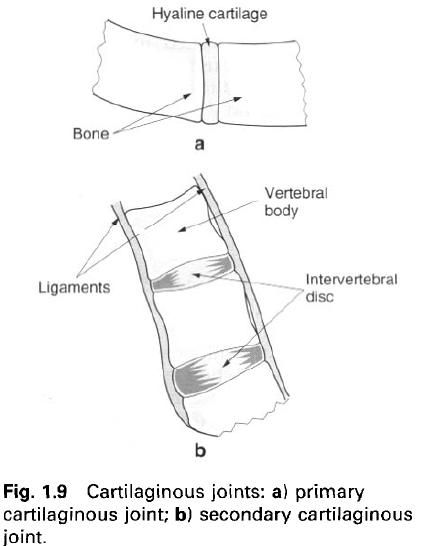
Secondary
cartilaginous(figure b)
These joints occur in the midline of the body
and are slightly more specialized. Moreover, their structure enables a small
amount of controlled movement to take place. Hyaline cartilage covers the
articular surfaces of the bones involved in the joint, but interposed between
these hyaline coverings is a pad of fibrocartilage. Examples are the joints
between the bodies of adjacent vertebrae, where the fibrocartilaginous pad is
in fact the intervertebral disc, and the joint between the two bodies of the
pubic bones.
Synovial joints
Synovial joints are a class of freely mobile
joints, with movement being limited by muscles, ligaments and the associated
joint capsules. The majority of the joints of the limbs are synovial. In
synovial joints are articular surfaces of the bones involved are covered with
articular(hyaline) cartilage, which because of its hardness and smoothness
enable the bones to move against each other with minimum friction. Passing
between the two bones, either attaching at or away from the articular margins,
is a fibrous articular capsule. The capsule my be strengthened by the blending
of ligaments or the deeper parts of muscles crossing the joint. Lining the deep
surface of the capsule is the synovial membrane, which covers all the surfaces
within the capsule except the articular cartilage(figure a). The synovial
membrane secretes synovial fluid into the joint space(cavity) enclosed by the
capsule, and serves to lubricate and nourish the articular cartilage as well as
the opposing joint surfaces. During movement the joint surfaces either glide or
roll past each other.
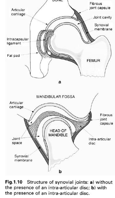
If the bones involved in the articulation
originally ossified in membrane, then the articular cartilage has a large
fibrous element. In addition there is enclosed within the capsule an
intra-articular disc, which may not be complete(figure b).
Bursae are often associated with synovial
joints, sometimes communicating directly with the joint space.
Because of the large number of synovial joints
within the body and their differing forms they can be subdivided according to
the shape of their articular surfaces and the movements possible at the joint.
Plane
joint
The joint surfaces are flat, or at least
relatively flat, and of approximately equal extent. The movement possible is of
a single gliding type or a twisting of one bone against the other, usually
within narrow limits. An example is the acromioclavicular joint.
Saddle
joint
The two surfaces are reciprocally
concavoconvex, as a rider sitting on a saddle. The principal movements possible
at the joint occur about two mutually perpendicular axes. However, because of
the nature of the joint surfaces there is usually a small amount of movement
about a third axis. The best example in the body is the carpometacarpal joint
of the thumb.
Hinge
joint
The surfaces are so arranged to allow movement
about one axis only. Consequently, the “fit” of the two articular surfaces is
usually good, but in addition the joint is supported by strong collateral
ligaments. The elbow is a typical hinge joint. The knee joint is considered to
be a modified hinge joint, as it permits some movement about a second axis. In
this case the movement is possible because of the poor fit of the articular
surfaces.
Pivot
joint
Again movement occurs about a single axis, with
the articular surfaces arranged so that one bone rotates within a fibro-osseous
ring. The atlantoaxial joint is a good example of a pivot joint.
Ball
and socket joint
As the name suggests the “ball” of one bone
fits into the “socket” of the other. This type of joint allows movement about
three principal mutually perpendicular axes. The hip joint is a ball and socket
joint.
Condyloid
joint
This is a modified form of a ball and socket
joint, which only allows active movement to occur about two perpendicular axes.
However, passive movement may occur about the third axis. The metacarpophalangeal
joints are examples of such joints.
Ellipsoid
joint
This is another form of a ball and socket
joint, although in this case the surfaces are ellipsoid in nature.
Consequently, movement only occurs about two perpendicular axes. The
radiocarpal joint is an ellipsoid joint.


0 коментара:
Постави коментар