Free Facebook Likes, Youtube Subscribers,  Twitter Followers

Ads 468x60px

Blogger Tricks

Blogger Themes

28. 2. 2013.

Lymphatics of the upper body


The lymphatic drainage of the upper limb is by a superficial intermeshing network of vessels just below the skin, and by deep lymphatic channels below the deep fascia. The larger lymph vessels contain numerous valves which allow lymph to move only in a proximal direction. Both groups of vessels drain proximally and end by passing through some of the 25 to 30 lymph nodes in the axilla. This mass of lymph nodes serves to filter the lymph contained in the system and acts as an important defence mechanism in preventing the spread of infection.
The axillary lymph nodes are distributed in the axillary fat throughout the axilla but can be divided into five groups, four of which lie below pectoralis minor and one(the apical group) above pectoralis major. Ultimately, all lymph from the upper limb passes through the apical group of nodes, from where the efferent lymph channels condense to form the subclavian trunk. On the left hand side, the subclavian trunk joins the thoracic duct, while on the right it drains into the subclavian vein directly or via the right lymphatic duct.

The superficial nodes and lymph vessels

The superficial lymph vessels are found in the skin and drain lymph from the superficial tissues. In the hand a fine meshwork of vessels exists, which drain into progressively larger channels as they pass up the arm. The only superficial lymph vessels which have any consistent course are the larger ones which follow the major superficial veins. These end by passing into the axilla.
In the cubital fossa one or two lymph nodes lie medial to the basilic vein, receiving lymph from the medial fingers and ulnar half of the hand and forearm. These are also one or two lymph nodes in the infraclavicular fossa associated with the cephalic vein. These receive vessels from the shoulder and breast. A single node may be found in the deltopectoral groove.

The deep nodes and lymph vessels

The deep lymph vessels of the upper limb are less numerous than the superficial vessels with which they have many connections. Lying below the deep fascia, they accompany the major arteries in the arm. Most pass directly to the lateral group of axillary nodes. Small nodes may occur along both the radial and ulnar arteries and deep within the cubital fossa. Efferents from all of these nodes lie alongside the axillary vein, and from the pectoral and subscapular nodes pass along the lateral thoracic and subscapular arteries respectively. A central group of nodes is formed above the floor of the axilla. Although all the group receives lymph from all areas, the main drainage of the limb is to the lateral group. Drainage of the breast and anterior chest wall is to the pectoral nodes, that of the scapular region and upper back is to the subscapular nodes. Efferents from these groups pass to the central and then the apical group of nodes, the latter also receiving efferents from the superficial infraclavicular nodes.

Application

The fact that the larger lymph vessels contain valves is important during massage techniques aimed at reducing oedema. The massage strokes are applied from distal to proximal, ending at the axilla, with sufficient depth to compress the lymph vessels and encourage drainage.
Active muscle contraction will also cause compression of the lymph vessels and encourage drainage proximally. This effect can be further enhanced by placing an elastic compressive support or bandage on the upper limb and encouraging active rhythmical contraction of the arm muscles. Elevation of the arm above the level of the axilla will allow gravity to assist the lymphatic drainage.
Pneumatic splints which apply a rhythmically alternating compressive force to the arm, using a small electric compressor pump, can also achieve the effect of increasing lymphatic flow proximally, utilizing the same principles as massage.

27. 2. 2013.

The veins of the upper body


The veins of the upper body

The veins of the upper limb are divided into a superficial group which lie in the superficial fascia, and a deep group which accompany the arteries. Both groups of veins have valves which allow proximal drainage (a fact which should be borne in mind when using massage) and drain into the axillary vein.


The deep veins

Apart from the axillary artery, which is accompanied by a single vein, all of the arteries are accompanied by two venae comitantes.
The axillary vein is the continuation of the basilic vein at the lower border of teres major. It ends by becoming the subclavian vein  at the lateral border of the first rib. Its course is identical to that of the axillary artery, which lies lateral to it.

The superficial veins

These are arranged in irregular networks in the superficial fascia. They are connected with the deep veins by inconstant perforating veins which pierce the deep fascia. The blood is drained from the superficial system principally by the basilica and cephalic veins.
The main superficial channels are as follows:

1) The dorsal venous arch

This would be better named the dorsal venous plexus, for its arch-like nature is seldom apparent. It lies on the back of the hand, its position and pattern being highly variable.

2) The basilic vein

This arises from the ulnar side of the arch and ascends along the ulnar side of the distal half of the forearm befoe inclining forwards to pass in front of the medial epicondyle and enetering the medial bicipital furrow. Opposite the insertion of coracobrachialis, it pierces the deep fascia to ascend along the medial side of the brachial vessels to become the axillary vein at the lower border of teres major. In the forearm it can usually be clearly seen, particularly in males. It is joined by tributaries from the forearm and by the median cubital vein in front of the elbow.

3) The cephalic vein

This arises from the radial end of the dorsal venous arch and receives the dorsal veins of the thumb. Inclining forwards, it ascends on the anterolateral part of the forearm as far as the elbow, and then along the lateral side of the biceps tendon to reach the groove in front of the shoulder between deltoid and pectoralis major(the deltopectoral groove). It ascends in this groove to the level of the coracoid process where it turns medially between pectoralis major and pectoralis minor. It pierces the clavipectoral fascia and ends in the axillary vein at a point just below the middle of the clavicle. It receives several tributaries in the forearm, and at the elbow is connected to the basilic vein by the median cubital vein.

4) The medial cubital vein

This is a short, wide vein, useful for venepuncture. It runs upwards and medially across the bicipital aponeurosis. The latter separates it from the underlying brachial artery. It joins the basilic vein just above the medial epicondyle.

5) The anterior median vein of the forearm

When present, this vein runs up the middle of the front of the forearm and may join the basilic or cephalic vein, or may divide at the cubital fossa into the median cephalic and median basilic veins. 

The arteries and pulses of the upper body


The main arterial stem of the upper limb passes through the root of the neck, the axilla and the arm before dividing into two in the forearm. It changes its name in each of the regions as it crosses particular bony or muscular landmarks. 


The subclavian artery

The right subclavian artery lies entirely within the root of the neck, having arisen from the brachiocephalic trunk. The left subclavian artery arises from the aortic arch in the superior mediastinum to enter the root of the neck. Both arteries pass laterally over the first rib towards the axilla, and end by becoming the axillary artery its lateral border. The subclavian artery is conveniently divided into three parts by scalenus anterior which crosses it anteriorly.
In the neck, the artery runs from the upper border of the sternoclavicular joint to the middle of the clavicle. The course is convex upwards. The artery can be compressed against the first rib by a downward and backward pressure applied behind the clavicle, lateral to the posterior border of sternocleidomastoid.
Branches from the subclavian artery supply structures within the neck, and the anterior chest wall, and give an important supply to the brain via the vertebral artery.

The axillary artery

The axillary artery is the continuation of the subclavian artery at the lateral border of the first rib and ends by becoming the brachial artery at the lower border of teres major, at the level of the lateral extremity of the posterior axillary fold. For descriptive purposes, it is divided into three parts by pectoralis minor. However, the length of each part will depend on the position of the arm. The cords of the brachial plexus are named according to their position with respect to the second part of the axillary artery.
The course of the artery is represented by a line drawn from the midpoint of the clavicle and passing immediately below the coracoid process to the medial lip of the intertubercular groove behind coracobrachialis. It thus describes a curve with the concavity facing downwards and medially.
The pulse of the axillary artery is readily palpated in the lateral wall of the axilla in the groove behind the coracobrachialis muscle. This is a useful pressure point to control distal bleeding, although paraesthesia may result from the inevitable pressure on the median, ulnar and radial nerves which are in close relation to the artery at this point.
Branches from the axillary artery supply the shoulder and pectoral regions, as well as the lateral chest wall. They anastomose with branches from the subclavian artery and the posterior intercostal arteries from the descending thoracic aorta. These anastomoses and their vessels may be enlarged in conditions where there is a narrowing of the aorta beyond the origin of the left subclavian artery(coarctation of the aorta), and serve as a collateral system bypassing the restriction.

The brachial artery

This is the continuation of the axillary artery beyond the lower border of teres major and ends in the cubital fossa opposite the neck of the radius. It lies successively on the long and medial heads of triceps, the coracobrachialis insertion and brachialis. Anteriorly it is covered by the medial border of the biceps, and is crossed from lateral to medial by the median nerve about halfway down the arm. In the cubital fossa it lies beneath the bicipital aponeurosis, which separates it from the median cubital vein, with the median nerve lying medial to the artery and with the biceps tendon lying lateral. The brachial artery divides into the radial and ulnar arteries.
High division of the brachial artery sometimes occurs proximal to the cubital fossa. Indeed, it may divide at any point between the axilla and the cubital fossa, in which case the two arteries descend side-by-side following the normal course of the brachial artery.
The brachial pulse may be felt along the whole course of the artery by compressing it against the humerus, directing lateral pressure proximally and dorsolateral pressure distally. It is best felt just medial to the bicipital aponeurosis at the level of the medial epicondyle of the humerus, and it is at this point that one listens for Korotkoff’s sounds when measuring blood pressure.
A major branch of the brachial artery is the profunda brachii, which runs with the radial nerve in the spiral groove between the lateral and medial heads of the triceps to pass into the posterior compartment of the arm. Branches from both the brachial artery and the profunda brachii supply the muscles of the arm and contribute to the anastomosis around the elbow joint.

The radial artery

The radial artery begins in the cubital fossa opposite the neck of the radius and ends by completing the deep palmar arch in the hand. It is usually thought of and described as having three parts. The first part is in the forearm, the second curves laterally around the wrist as far as the first interosseus space, and the third passes through the interosseus space into the palm.
If the arm is placed in a midpronated position and brachioradialis is tensed, then the course of the first part of the radial artery may be indicated by a slightly convex line beginning at the biceps tendon and running down the medial side of brachioradialis to a point just medial to the radial styloid process on the anterior aspect. As it curves around the wrist, the radial artery is within the “anatomical snuffbox” and lies on the radiocarpal ligament, the scaphoid and trapezium. It is crossed by the tendons of abductorpollicis longus and extensors pollicis brevis and longus from lateral to medial.
The third part of the artery passes between the two heads of the first dorsal interosseus and adductor pollicis before completing the deep palmar arch.
The radial pulses may be felt against the distal border of the radius lateral to flexor carpi radialis, and in the “anatomical snuffbox” against the scaphoid.
Branches from the first part of the artery are involved in the elbow anastomosis and supply the muscles on the radial side of the forearm. From the second part arise branches supplying the wrist and dorsum of the hand and thumb. Before completing the deep palmar arch, the third part of the artery gives off the princes pollicis and radialis indicis branches to the thumb and index fingers respectively.

The ulnar artery

The ulnar artery begins in the cubital fossa as a terminal branch of the brachial artery, and ends at the pisiform by dividing into deep and superficial palmar arteries. It may be represented by a line passing, medially convex, from the tendon of biceps to the pisiform bone and from there to the hook of the hamate. In its course, it lies on brachialis, flexor digitorum profundus and the flexor retinaculum, and is crossed anteriorly, from above downwards by: pronator teres, the median nerve, flexor carpi radialis, palmaris longus and flexor digitorum superficialis, being overlapped lower down by flexor carpi ulnaris. Just below the radial tuberosity, the common interosseus artery is given off, and this divides into anterior and posterior interosseus arteries which run down either side of the interosseus membrane, supplying the deep muscles of both flexor and extensor compartments. Branches from the proximal and distal ends of the artery are involved in the supply to the elbow and wrist joints respectively.

The superficial palmar arch

This is formed mainly by the ulnar artery, with a contribution from the superficial palmar branch of the radial artery. It lies deep to the palmar aponeurosis. The distal convexity of the arch lies level with the flexor surface of the extended thumb.
Four common palmar digital arteries arise from the superficial arch with the most medial running along the medial side of the little finger. The other three each divide into two proper digital arteries which supply adjacent sides of the little, ring, middle and index fingers.

The deep palmar arch

This is formed mainly by the radial artery with a contribution from the deep branch of the ulnar artery. It lies deep to the long flexor tendons and their synovial sheaths on the bases of the metacarpals and gives rise to the palmar metacarpal arteries. Its distal convexity is 2cm distal to the distal crease of the wrist.

26. 2. 2013.

Dermatomes of the upper limb


During development cells from the dermomyotome spread out to form the skin. The dermatome represents an area of skin innervated by a single nerve root. Figure shows the dermatomes of the upper limb. Obviously branches from more than one nerve can and do contribute an individual dermatome, however, all will have the same single root value. 


Plexus brachialis - part III


The radial nerve

The radial nerve is the major nerve from the posterior cord, root value C5, 6, 7, 8 (T1), being one of its terminal branches. In the axilla, the radial nerve lies behind the axillary and upper part of the brachial arteries, passing anterior to the tendons of subscapularis, latissimus dorsi and teres major. The radial nerve, together with the profunda brachii artery, enters the posterior compartment of the arm by passing through the lower triangular space, formed by the humerus laterally, the long head of triceps medially and teres major above. In passing through this space, the nerve enters the spiral(or radial) groove of the humerus, descending obliquely between the lateral border of the humerus in the distal third of the arm. The nerve pierces the lateral and medial heads of triceps, reaching the lateral border of the humerus in the distal third of the arm. The nerve pierces the lateral intermuscular septum to enter the anterior compartment where it lies in a muscular groove between brachialis and brachioradialis. In front of the lateral epicondyle of the humerus, the radial nerve divides into its terminal superficial and deep branches.


In the arm, the radial nerve gives a supply to all three heads of triceps, anconeus, the lateral part of brachialis, brachioradialis and extensor carpi radialis longus. The branches to triceps all arise before the radial nerve enters the spiral groove; anconeus is supplied by a branch to the medial head of triceps.
The radial nerve also gives articular branches to the elbow joint and has three cutaneous branches which supply skin on the back of the arm and forearm(figure a).
The posterior cutaneous nerve of the arm arises in the axilla, piercing the deep fascia near the posterior axillary fold. It supplies skin on the posterior surface of the proximal third of the arm.
The lower cutaneous nerve of the arm arises before the radial nerve pierces the lateral intermuscular septum, and becomes cutaneous just below deltoid. It supplies the skin over the lower lateral part of the arm and a small area on the forearm.
The posterior cutaneous nerve of the forearm arises just below the previous nerve, and supplies a variable area of skin on the dorsum of the forearm as far as the wrist, or occasionally beyond.
The superficial branch is the direct continuation of the radial nerve, beginning in front of the lateral epicondyle and descending along the anterolateral side of the forearm. It is entirely sensory. It lies on supinator, pronator teres, flexor digitorum superficialis and flexor pollicis longus covered by brachioradialis with the radial artery medial to it. In the distal third of the forearm, the nerve passes below the tendon of brachioradialis and pierces the deep fascia to become superficial. It supplies the skin on the dorsum of the wrist, the lateral dorsal surface of the hand and dorsum of the thumb, and then divides into four or five digital nerves. The digital nerves supply the skin on the dorsum of the thumb, index, middle and adjacent half of the ring finger as far as the distal interphalangeal joint. The digital branches also give articular branches to the metacarpophalangeal and proximal interphalangeal joints of all five digits.
The deep branch, more often called the posterior interosseus nerve, is entirely muscular and articular. It begins in front of the lateral epicondyle of the humerus and enters the posterior compartment of the forearm by passing between the two heads of supinator, thereby curving around the lateral and posterior surfaces of the radius. During its course, the nerve supplies both extensor carpi radialis brevis and supinator. It then descends between the deep and superficial groups of extensor muscles, accompanied by the posterior interosseus artery, supplying all the muscles in the extensor compartment of the forearm: extensor digitorum, extensordigiti minimi, extensor carpi ulnaris, extensor pollicis longus, extensorindicis, abductor pollicis longus and extensor pollicis brevis.
In the lower part of the forearm, the posterior interosseus nerve lies on the interosseus membrane and ends in a flattened expansion, which gives articular branches to the intercarpal joints.

Applied anatomy

The radial nerve is often injured as it crosses the humerus, either as the result of a fracture or by pressure from a direct blow or incorrect use of a crutch. Triceps usually escapes denervation as it derives its supply from branches given off high in the arm, but a total paralysis of the extensors of the wrist and digits leads to the deformity of a “dropped wrist”(figure a). As a result, any attempt to grip or make a fist leads to increased flexion of the wrist and an inability to carry out effective movement. This is due to the loss of the synergic action of the wrist extensors which usually prevent the unwanted flexion of the wrist produced by the continued action of the finger flexors.


The interphalangeal joints of the fingers can be extended by the lumbricals and interossei which have an attachment to the dorsal digital expansion, but proper use of the hand requires effective form of “lively” splint which compensates for the paralysed muscles. Even though the sensory distribution of the radial nerve on the dorsum of the hand appears extensive, overlap by adjacent cutaneous nerves means that the area of exclusive radial nerve supply is a small patch on the dorsum of the thumb web.

The median nerve

The median nerve is complex in that it arises partly from the lateral cord (C5, 6, 7) and partly from the medial cord (C8, T1) of the brachial plexus. These two contributing heads of the median nerve unite by embracing the third part of the axillary artery. Once formed, the nerve descends under cover of biceps passing at first laterally to the brachial artery and then medially, having crossed it anteriorly. In the lower part of the arm the median nerve lies on brachialis, and in the cubital fossa is protected by the bicipital aponeurosis which crosses it.
The median nerve enters the forearm by passing between the two heads of pronator teres, and then runs below the tendinous arch connecting the heads of flexor digitorum superficialis to gain access to its deep surface. Closely bound to the deep surface of flexor digitorum superficialis, it descends on flexor digitorum profundus until just above the wrist where it becomes superficial by passing between the tendons of flexor digitorum superficialis and flexor carpi radialis, deep to palmaris longus. The median nerve enters the hand deep to the flexor retinaculum, passing anteriorly to the long flexor tendons. Consequently, it is one fo the structures found within the carpal tunnel.
During its course the median nerve gives articular branches to the elbow joint and supplies pronator teres, flexor carpi radialis, palmaris longus and flexor digitorum superficialis.
The palmar cutaneous nerve arises in the distal third of the forearm. It pierces the deep fascia and enters the palm by passing superficial to the flexor retinaculum. It supplies a small area of skin on the lateral side of the palm and thenar eminence.
In the cubital fossa, the anterior interosseus nerve arises from the median nerve and descends, with the anterior interosseus artery, on the anterior surface of the interosseus membrane between flexor pollicis longus and flexor digitorum profundus. It then runs deep to pronator quadratus eo end at the wrist by giving articular branches to the radiocarpal and intercarpal joints. The anterior interosseus nerve supplies flexor pollicis longus, the lateral half of flexor digitorum profundus and pronator quadratus.
Once the median nerve has passed through the carpal tunnel to enter the hand, it divides into lateral and medial terminal branches. The lateral branch passes laterally and proximally to enter the thenar eminence and supply abductor pollicis brevis, flexor pollicis brevis, opponens pollicis and the first lumbrical. It gives sensory branches to the adjacent sides of the thumb and index finger.
The medial branch of the median nerve divides into a variable number of branches, the palmar digital nerves, the most lateral of which supplies the second lumbrical. These nerves are sensory to the palmar surface of adjacent sides of the index and middle, and middle and ring fingers(figure a). Each of these digital nerves produces a dorsal branch which passes backwards to supply the dorsal aspect of the distal phalanx and nail bed, and a variable amount of the middle phalanx of the same digits.


The digital nerves lie deep to the palmar aponeurosis and superficial palmar arch, but superficial to the long flexor tendons. As well as the sensory innervation, they also give articular branches to the interphalangeal and metacarpophalangeal joints.

Applied anatomy

The median nerve can be injured in the forearm by deep cuts with a resultant loss of flexion at all interphalangeal joints, except the distal ones in the ring and little fingers. The metacarpophalangeal joints of these same fingers can still be flexed by the lumbricals and interossei but the movement of pronation is severely restricted. In the hand, the thumb is held in extension and adduction thus losing its ability to oppose and abduct. This, combined with the sensory loss, proves a major disability. More commonly the nerve is damaged just proximal to the flexor retinaculum by laceration, or deep to it in the carpal tunnel where compression gives rise to the carpal tunnel syndrome. In this instance only the thenar muscles, lateral two lumbricals and sensation in the hand will be affected. 


22. 2. 2013.

Plexus brachialis - part II


The axillary nerve

The axillary nerve arises from the posterior cord of the brachial plexus and has a root value of C5, 6. In the axilla it descends behind the axillary artery and in front of subscapularis, at the lower border of which it passes backwards close to the inferior part of the shoulder joint in company with the posterior circumflex humeral vessels. It then passes through the quadrilateral space where it supplies the shoulder joint and divides into anterior and posterior branches. The anterior branch winds around the surgical neck of the humerus, deep to and as far as the anterior part of deltoid, which it supplies. The posterior branch supplies teres minor, and the posterior part of deltoid. It then passes around deltoid as the upper lateral cutaneous nerve of the arm which pierces the deep fascia to supply skin over the lower part of deltoid and the lateral head of triceps as far as the middle part of the arm.


The axillary nerve is frequently injured when the shoulder is dislocated because of  its close proximity to this joint. Paralysis of deltoid and teres minor results, and consequently there is inability to abduct the arm beyond that possible by the action of supraspinatus. This, in conjuction with an area of anaesthesia over the back of deltoid and lateral head of triceps, allows a clinical diagnosis of nerve injury to be made.

The musculocutaneous nerve

The musculocutaneous nerve arises from the lateral cord of the brachial plexus and has a root value C5, 6, 7. It lies firstly lateral to the axillary artery and then descends between the artery and coracobrachialis which it supplies and pierces before running distally between biceps and brachialis to reach the lateral side of the arm. At the elbow, the musculocutaneous nerve pierces the deep fascia between biceps and brachioradialis as the lateral cutaneous nerve of the forearm.


In the arm the musculocutaneous nerve supplies both heads of biceps brachii and two-thirds of brachialis as well as coracobrachialis.
The lateral cutaneous nerve of the forearm divides into anterior and posterior branches. The anterior branch supplies the skin on the lateral half of the forearm as far as the ball of the thumb, while the posterior branch supplies a variable area over the extensor muscles of the forearm, wrist and occasionally the first metacarpal.

The ulnar nerve

The ulnar nerve is one of the terminal branches of the medial cord of the brachial plexus, having a root value C8 and T1, but frequently contains fibres from C7. It descends on the medial side of the axillary artery behind the medial cutaneous nerve of the forearm, and continues downwards medial to the brachial artery, anterior to triceps. In the distal half of the arm, the ulnar nerve passes backwards and pierces the medial intermuscular septum to enter the posterior compartment of the arm, where it lies on the front of the medial head of triceps. Continuing its descent in the posterior compartment of the arm, the ulnar nerve passes between the medial epicondyle of the humerus and the olecranon of the ulna, lying in the ulnar groove behind the medial epicondyle. The ulnar nerve then enters the anterior compartment of the forearm by passing between the two heads of flexor carpi ulnaris, initially in contact with the ulnar collateral ligament of the elbow. As it descends along the medial side of the forearm, the ulnar nerve lies on flexor digitorum profundus, lateral to the ulnar artery, covered in its upper part by the belly of flexor carpi ulnaris, but in the lower half, only by its tendon. Proximal to the flexor retinaculum it pierces the deep fascia to lie lateral to flexor carpi ulnaris, and passes anterior to the flexor retinaculum lateral to the pisiform where it divides into superficial and deep branches.


During its course, the ulnar nerve gives an articular branch to the elbow joint, and supplies flexor carpi ulnaris, and the medial half of flexor digitorum profundus.
A palmar cutaneous branch arises from the ulnar nerve piercing the deep fascia in the distal third of the forearm and descends to supply the skin over the medial part of the palm.
The dorsal branch of the ulnar nerve also arises in the distal third of the forearm, passes backwards deep to flexor carpi ulnaris to pierce the deep fascia on the medial side to become superficial. On the medial side of the wrist, it crosses the triquetral, against which it can be palpated, and gives branches to the dorsal surface of the wrist and hand. Here it divides into two or three dorsal digital nerves which supply the skin on the dorsum of the hand and the dorsal surfaces of the medial one and a half or two and a half fingers, excluding the skin over the distal phalanx. The dorsum of the distal phalanges is supplied by branches from the median or ulnar nerves derived from the palm.
The superficial branch of the ulnar nerve lies deep to palmaris brevis on the medial side of the hand where it can be compressed against the hook of the hamate. It supplies the palmaris brevis muscle, the skin on the medial side of the palm of the hand, and the skin on the palmar surface of the little and adjacent half of the ring fingers, extending onto the dorsal surface supplying the skin and nail bed of the distal phalanx.
The deep branch of the ulnar nerve eventually runs with the deep branch of the ulnar artery and thus loops across the palm from medial to lateral deep to the flexor tendons. It passes initially between abductor digiti minimi and flexor digiti minimi and pierces opponens digiti minimi, supplying all three muscles. As it passes across the deep part of the palm, it supplies the medial two lumbricals, all of the interossei and adductor pollicis. Rarely, the ulnar nerve also supplies the thenar muscles. The deep branch gives articular filaments to the wrist joint.

Applied anatomy

The ulnar nerve may be damaged in the groove behind the medial epicondyle either by trauma or entrapment. This leads to a partial of complete loss of muscular and sensory innervation. At the wrist, the nerve can easily be cut or lacerated because of its superficial position. The clinical picture can be complicated if the lesion occurs below the level where the dorsal and palmar cutaneous branches are given off, as a considerable portion of the skin on the ulnar side of the hand still has a sensory supply. The result of an ulnar nerve lesion often gives the typical “claw-hand deformity”.


This is due to loss of power in the intrinsic muscles of the hand and the unopposed actions of antagonistic muscle groups. There is “guttering” between the metacarpals, an inability to abduct the fingers or adduct the thumb. The area of sensory loss usually follows the outline of the sensory map.


21. 2. 2013.

Plexus brachialis - part I


Introduction

The nerves supplying the structures in the arm are all derived from the brachial plexus, a complex of intermingling nerves originating in the neck.


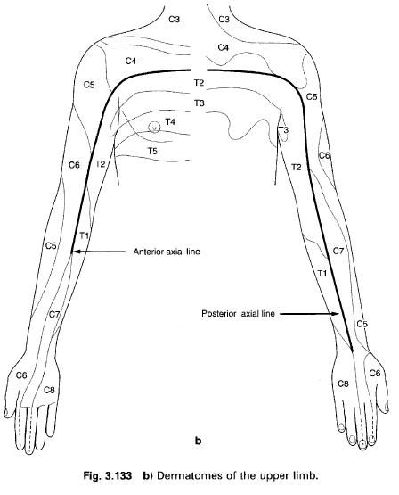
The brachial plexus is formed by the ventral rami of the lower four cervical nerves and the first thoracic nerve to give it a root value of C5, 6, 7 and 8 and T1. Occasionally, there may be a contribution from C4 or T2 or both.
The ventral rami are found as the anterior division of the spinal nerve, just outside the intervertebral foramen, lying between scalenus anterior and medius. They are collectively termed the roots of the plexus. Each spinal nerve receives an autonomic contribution, C5 and 6 receiving grey rami communicates from the middle cervical ganglion, while C7, 8, and T1 receive them from the inferior or cervicothoracic ganglion.
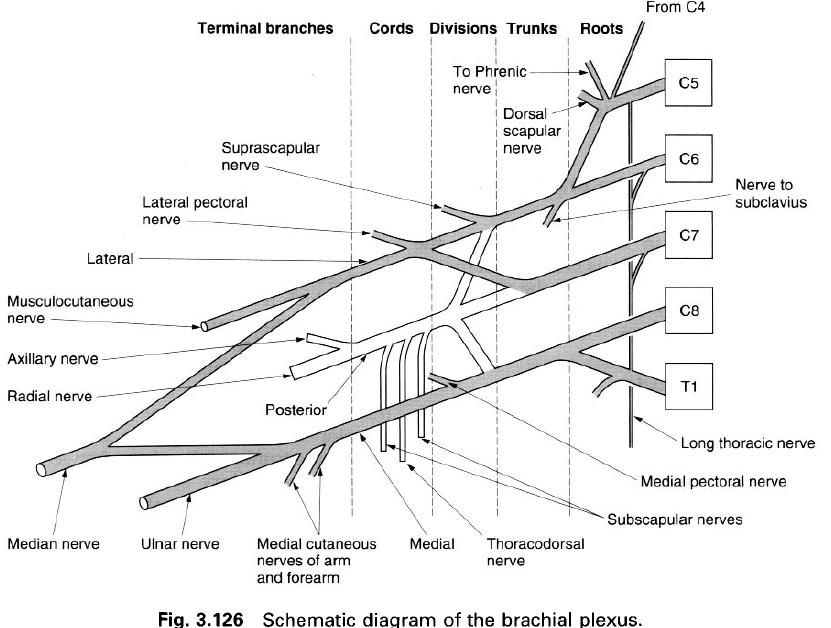
Commonly, the upper two roots(C5 and 6) unite to form the upper trunk, the lower two roots(C8 and T1) unite to form the lower trunk, and the C7 root continues as the middle trunk. These three trunks are found running between the scalene muscles and the upper border of the clavicle in the posterior triangle of the neck. The lower trunk may groove the upper surface of the first rib behind the subclavian artery; the T1 root is always in contact with the rib.
Just above the clavicle, each of the three trunks divides into an anterior and posterior division which supply the flexor and extensor compartments of the arm respectively. The three posterior divisions unite to form the posterior cord, while the anterior divisions of the upper and middle trunks unite to form the lateral cord, and the anterior division of the lower trunk continues as the medial cord. These three cords pass downwards into the axilla, running firstly posterolateral to the axillary artery, but then in their named positions with respect to the second part of the axillary artery posterior to pectoralis minor, that is medial, posterior and lateral. The cords and axillary artery are bound together in an extension of the prevertebral fascia which protrudes into the axilla, the axillary sheath.

Applied anatomy

The brachial plexus itself is subject to direct injury, consequently a knowledge of its formation is of help in determining in exactly which parts, and at what levels, the damage has occurred. Traction injuries occur when the roots of the plexus are torn from the spinal cord, or when the constituent parts are partially or completely torn. In extreme cases, the whole plexus may be disrupted to produce a completely denervated arm. If the upper roots are completely torn, and Erb’s paralysis affecting the musculature of the upper arm is produced; whereas if the lower roots are completely torn, a Klumpikes’ paralysis affecting the hand and forearm will result.

Nerves arising from the brachial plexus and their distribution

The simplest way in which to describe the nerves of the brachial plexus is to put them into terminological order relating to the part of the plexus from which they originate(and to indicate their root value).

Branches from the roots

  1. Nerves to the scalene and longus colli muscles(C5, 6, 7, 8).
  2. A branch to the phrenic nerve(C5).
  3. The dorsal scapular nerve(C5).
  4. The long thoracic nerve(C5,6,7).

Branches from the trunks

  1. The nerve to subclavius muscle(C4, 5, 6).
  2. Suprascapular nerve(C4, 5, 6).

There are no nerves arising from the divisions.

Branches from the cords of the plexus

Medial cord:

  1. Medial pectoral nerve(C8, T1).
  2. Medial cutaneous nerve of the forearm(C8, T1).
  3. Medial cutaneous nerve of the arm(T1).
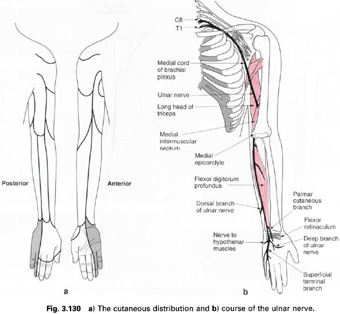
  4. Ulnar nerve(C7, 8, T1).
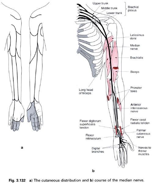
  5. Medial part of the median nerve(C8, T1).

Posterior cord:

  1. Upper subscapular nerve(C4, 5, 6, 7).
  2. Thoracodorsal nerve(C6, 7, 8).
  3. Lower subscapular nerve(C5, 6).
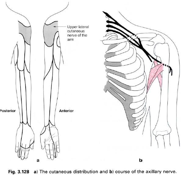
  4. Axillary nerve(C5, 6).
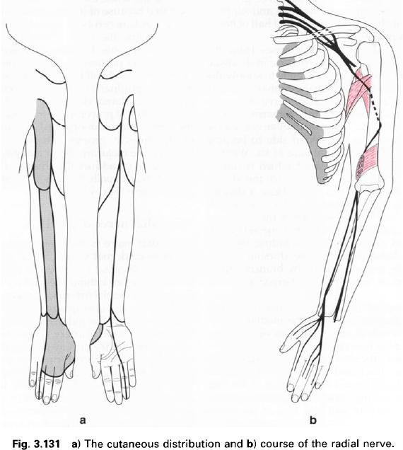
  5. Radial nerve(C5, C6, C7, C8, T1).

Lateral cord:

  1. Lateral pectoral nerve(C5, 6, 7).
  2. Musculocutaneous nerve(C5, 6, 7).
  3. Lateral part of the median nerve(C5, 6, 7).

Branches from the roots

The muscular supply to the scalene and longus colli muscles arises by twigs from the upper surface of the anterior primary rami as they emerge from the intervertebral foramina, directly entering the muscles. The C5 contribution to the phrenic nerve arises as the lateral border of scalenus anterior.
The dorsal scapular nerve(C5) passes through scalenus medius to gain the deep surface of the levator scapulae muscle, from which it runs onto the anterior surface of the rhomboid muscles. It supplies both rhomboid major, minor and levator scapulae.
The C5 and 6 roots of the long thoracic nerve unite after piercing scalenus medius and are joined by the C7 root on the anterior surface of the muscle. The nerve passes behind the trunks of the plexus between the first rib and the axillary artery to gain the outer(axillary) surface of serratus anterior which it supplies. The upper two digitations of the muscle are supplied by C5, the next two by C6, and the remaining four by C7.
The long thoracic nerve may be damaged by direct pressure on it from above the shoulder.
The resultating paralysis of serratus anterior causes the characteristic “winged” scapula, with the inability to perform activities, such as abduction of the arm, where the scapula is stabilized or laterally rotated.

Branches from the trunks

The nerve to subclavius(C4, 5, 6) is a small branch from the upper trunk. It descends anterior to the subclavian artery to supply subclavius. It may communicate with the phrenic nerve.
The suprascapular nerve(C4, 5, 6) is a large branch from the upper trunk. It passes inferolaterally above and parallel to the trunks through the suprascapular notch deep to trapezius to enter the supraspinous fossa of the scapula. It then runs deep to the supraspinatus muscle and enters the infraspinous fossa via the spinoglenoid notch. The suprascapular nerve supplies both supraspinatus and infraspinatus, and also gives articular filaments to the shoulder and acromioclavicular joints.
All of the above branches arise from the plexus above the clavicle, whereas those considered below all arise below the level of the clavicle in the axilla.

Branches from the cords

The lateral pectoral nerve arises from the lateral cord of the plexus with a root value of C5, 6, 7. It crosses anteromedially in front of the axillary artery, giving a branch to the medial pectoral nerve before piercing the clavipectoral fascia to gain access to the deep surface of pectoralis major which it supplies.
The medial pectoral nerve arises from the medial cord with a root value C8, T1. It receives a contribution from the lateral pectoral nerve and passes between the axillary artery and vein to gain access to the deep surface of pectoralis minor which it supplies. It then pierces this muscle to end in pectoralis major which it also supplies.
The upper and lower subscapular nerves both arise from the posterior cord, root values C4, 5, 6, 7 respectively. From behind the axillary artery they descend towards the subscapular fossa where they both supply the subscapularis muscle. In addition, the lower subscapular nerve enters and supplies teres major.
The thoracodorsal nerve also arises from the posterior cord(C6, 7, 8), between the two subscapular nerves. The nerve passes inferomedially along the posterior wall of the axilla, and the anterolateral surface of latissimus dorsi before entering its deep surface to supply it.
The medial cutaneous nerve of the arm is a small nerve arising from the medial cord(root value T1). It descends through the axilla on the medial side of the axillary vein, and then along the medial side of the brachial artery. It pierces the deep fascia to supply the skin and fascia on the medial side of the proximal half of the arm, extending onto both the anterior and posterior surfaces. It may be partly or entirely replaced by the intercostobrachial nerve(root value T2, 3).
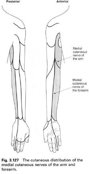
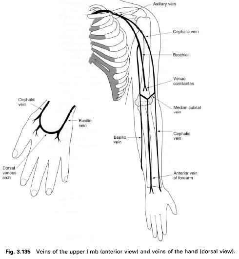
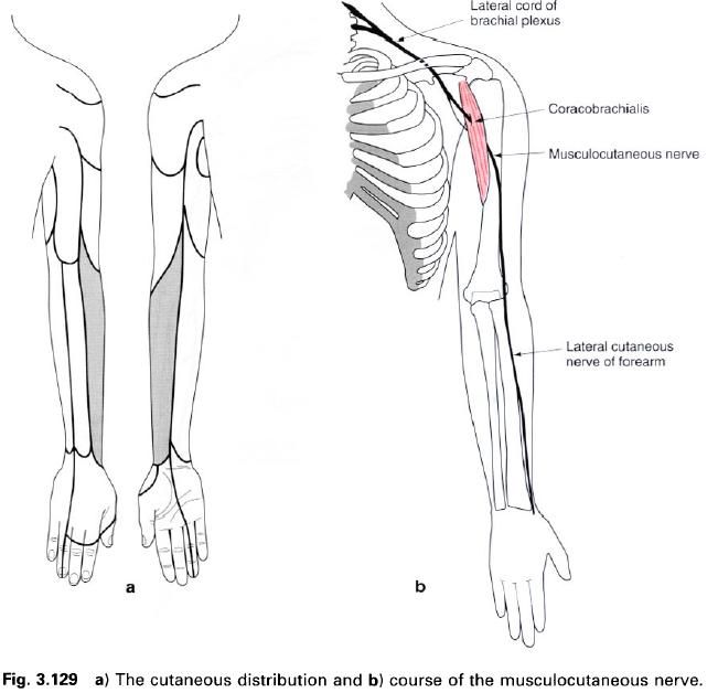


The medial cutaneous nerve of the forearm arises directly from the medial cord with a root value of C8, T1. It descends on the medial side of the axillary and brachial arteries. It pierces the deep fascia, together with basilic vein, in the middle of the arm, and descends with it to the elbow, where it then divides into anterior and ulnar branches. The medial cutaneous nerve of the forearm supplies the skin over the lower part of biceps, the medial side of the forearm as far as the wrist, and part of the medial side of the posterior surface of the forearm.


Search this blog