


Interphalangeal joints
Because each finger consists of three phalanges
it contains two interphalangeal joints: a proximal joint between the head of
the proximal and base of the middle phalanx, and a distal joint between the
head of the middle and base of the distal phalanx. All of the joints are hinge
joints permitting flexion and extension only, with the articular surfaces
covered by hyaline cartilage.
Articular
surfaces
The articulation is between the pulley-shaped
head of the phalanx and two shallow facets separated by a ridge on the base of
the immediately distal phalanx. The groove and ridge on the head and base
respectively do not lie exactly in an anteroposterior direction, except for the
joints of the index finger. In all other joints they run slightly obliquely
from posterolateral to anteromedial, with the obliquity increasing from the
middle to the little finger.
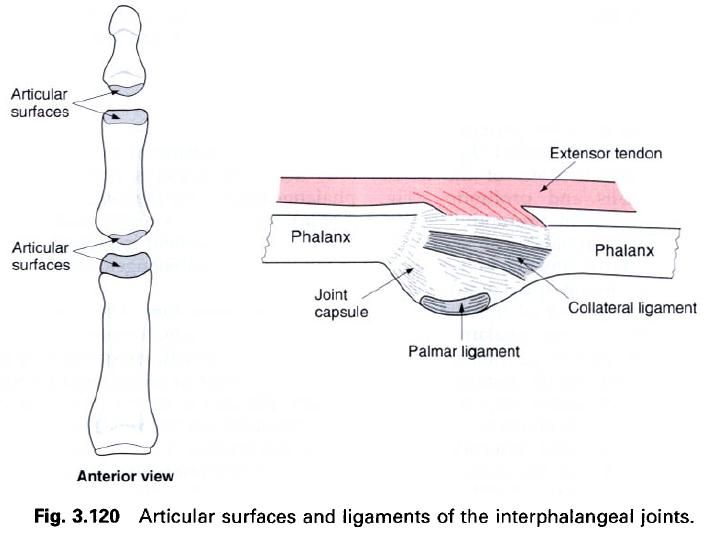
The articular surface of the phalangeal head is
greater than that on the adjacent base, extending further distally on its
anterior aspect. The head is also wider anteriorly than posteriorly. A fibrocartilaginous
plate, the palmar ligament, similar to that associated with the
metacarpophalangeal joint, acts as a mobile articulate surface.
Joint
capsule and synovial membrane
A loose fibrous capsule surrounds the joint,
being strengthened at the sides by collateral ligaments, and partly replaced
anteriorly and posteriorly by the palmar ligament and extensor expansion
respectively. Synovial membrane lines all non-articular surfaces, including the
anterior and posterior recesses of the capsule.
Ligaments
The collateral
ligaments attach to the sides of the head of the most proximal phalanx,
blending with the margins of the palmar ligament. They tend to be not so
obliquely orientated as the collateral ligaments of the metacarpophalangeal
joints. The ligaments become increasingly tense with flexion at the joint.
The palmar
ligament is a mobile fibrocartilaginous plate attached to the anterior
margin of the base of the adjacent phalanx. It is loosely attached to the front
of the neck of the immediately preceding phalanx by the joint capsule. Also
attached to the palmar ligament is the fibrous flexor sheath of the digit.
Blood
and nerve supply
The arterial supply to the joints is by
branches from the digital arteries running along the sides of each finger.
Anteriorly the digital arteries arise from the palmar metacarpal arteries,
while posteriorly they come from the dorsal metacarpal arteries. Venous
drainage is by similarly named vessels, eventually draining into the venae
comitantes associated with the radial and ulnar arteries. Posteriorly some
venous drainage will pass to the dorsal venous plexus on the dorsum of the
hand, and thence into the basilica and cephalic veins. Lymphatic drainage from
the joints is by vessels which follow the arteries, with the majority of lymph
draining to the lateral group of axillary nodes, although some may pass to
cubital or brachial nodes.
The nerve supply to the joint is by twigs from
adjacent digital nerves, and have a root value of C7. For the index, middle and
lateral side of the ring fingers, the digital nerves are branches of the median
nerve anteriorly and the radial nerve posteriorly. For the medial side of the
ring and the little fingers the digital nerves all arise from the ulnar nerve.
Relations

On the anterior aspect of the proximal
interphalangeal joint, enclosed within the fibrous flexor sheath, are the
tendons of the flexor digitorum superficialis and profundus(picture below); only the tendon of profundus lies in front of the distal
interphalangeal joint. The fibrous flexor sheaths are relatively thin and loose
over the interphalangeal joints, with a cruciate arrangement of fibres as they
pass from the side of one phalanx to the opposite side of the preceding
phalanx. Immediately beyond the distal interphalangeal joint, the flexor sheath
attaches to the palmar surface of the distal phalanx.
Posterior to the proximal interphalangeal joint
is the central slip of the dorsal digital expansion. On the back of the middle
phalanx the two collateral slips of the expansion come together, so that a
single tendon crosses the posterior aspect of the distal interphalangeal joint.
Stability
The interphalangeal joints are fairly stable
because of the presence of the long flexor and extensor tendons. Nevertheless dislocations
can and do occur; they can often be reduced by manipulation.
Movements
Because of the nature of the joint surfaces,
the only active movements possible at the interphalangeal joints are flexion
and extension(figure a). However, a small degree of passive side-to-side
movement is possible, particularly at the distal interphalangeal joint.
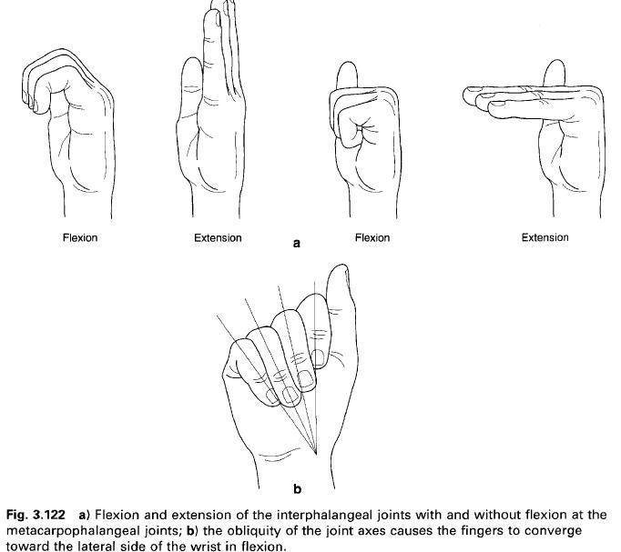
Flexion
and extension. These take
place about a transverse axis, which for the middle, ring and little fingers
with increasing obliquity from lateral and distal to medial and proximal(figure
b). The axis is approximately perpendicular to the groove on the phalangeal
head, so that when the medial fingers are flexed at the interphalangeal joints
the movement does not occur in a sagittal plane, but enables these fingers to
oppose the thumb more easily. Flexion of the index finger, however, occurs in a
sagittal plane.
The range of flexion at the proximal
interphalangeal joint is greater than 90° for all fingers, and gradually
increases towards the little finger so that this is capable of 135° of flexion.
At the distal interphalangeal joint, the range of flexion for little fingers is
90°, and gradually decreases towards the index finger. Active extension at the
interphalangeal joint is minimal, being no more than 5° at the distal and only
1° to 2° at the proximal joints. Passive extension may be considerably greater.
Flexion at the proximal interphalangeal joint
is primarily due to the action of flexor digitorum superficialis, assisted by flexor digitorum profundus. Only profundus
flexes the distal interphalangeal joint. Extension of the interphalangeal
joints is produced by contraction of the lumbrical
and interossei via their attachment to the dorsal digital expansions. They
are assisted in each finger by extensor digitorum, and in the index and little fingers by extensors indicis and digiti minimi respectively.
Simultaneous flexion at one interphalangeal
joint and extension at the other is produced by a controlled balance between
the activity of the flexor and extensor muscles. Flexion of the wrist
facilitates extension of the fingers and opening the fist. The functional
position of the wrist(that is in extension) puts the finger flexors beyond
their natural length and so enables greater tension to be developed in them,
facilitating a powerful grip. Similarly, flexing the interphalangeal joints
places the extensors of the wrist under increased tension. In stabilizing the
wrist, a certain amount of flexor strength amd extensor power is sacrified.
Only 70% of the strength of the finger flexors is available from flexion of the
interphalangeal joints. Weakness of the wrist extensors, by falling to maintain the position of function, greatly
interferes with the strength of the finger flexors and the ability to carry out a forceful closure of the fist.


0 коментара:
Постави коментар