


Extensor carpi radialis longus
Extensor carpi radialis brevis
Extensor carpi ulnaris
Extensor digitorum
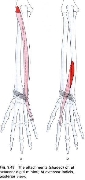
Extensor indicis
Extensor digiti minimi
Extensor pollicis longus
Extensor pollicis brevis
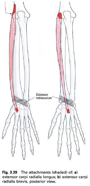
Extensor carpi radialis longus
Extensor carpi radialis longus lies on the
lateral side of the posterior compartment of the forearm, being partly covered
by brachioradialis. It arises from
the anterior part of the lower third
of the lateral supracondylar ridge of
the humerus and adjacent intermuscular septum. Occasionally, there may be an
attachment to the lateral epicondyle by the common extensor tendon.
Approximately in the middle of the forearm, the muscle forms a flattened tendon
which runs distally over the lateral surface of the radius. In the lower third of the forearm, the tendon, together
with that of extensor carpi radialis brevis, is crossed by the tendons abductor
pollicis longus and extensor pollicis brevis. The tendon of extensor carpi
radialis longus and brevis pass deep to the extensor retinaculum in a common
synovial sheath. Together they groove the posterior surface of the styloid
process of the radius. The tendon of
extensor carpi radialis longus attaches to the posterior surface of the base of
the second metacarpal.
Nerve
supply
Extensor carpi radialis longus is supplied by
the radial nerve, root value C6, 7, which enters the muscle above the elbow.
The skin over the muscle is supplied by roots C5, 6.
Action
and palpation
These will be considered with extensor carpi
radialis brevis.
Extensor carpi radialis brevis
Extensor carpi radialis brevis lies adjacent to
and is partly covered by longus, to which it may be partly fused. It arises
from the lateral epicondyle of the humerus via the common extensor tendon, the lateral
ligament of the elbow and the
adjacent fascia. The tendon of brevis forms halfway down the forearm and runs
with that of longus deep to abductor pollicis longus and extensor pollicis
brevis. It passes below the extensor retinaculum, in a common synovial sheath,
to attach to the posterior surface of
the base of the third metacarpal.
Nerve
supply
Extensor carpi radialis brevis is supplied by
the posterior interosseus branch of
the radial nerve, root value C6, 7.
The skin over the muscle is supplied by roots C5, 6, 7.
Action
Working with extensor carpi ulnaris, extensor carpi
radialis longus and brevis produce extension of the wrist. Working with the
flexor carpi radialis, however, they will produce abduction(radial deviation)
of the wrist. In addition, extensor carpi radialis longus may help to flex the
elbow joint.
Functional
activity
Functionally, the wrist extensors work strongly
in the action of gripping, where they have a synergic role. The synergy of the
two radial extensors and extensor carpi ulnaris is a vital factor in the
gripping action. By maintaining the wrist in an extended position, flexion of
the wrist under the action of flexor digitorum superficialis and profundus is
prevented, with the result that these muscles act on the fingers. If the wrist
is then allowed to flex the flexor tendons cannot shorten sufficiently to
produce effective movement at the interphalangeal joints. This therefore
becomes a state of active insufficiency.
If the radial nerve is damaged the patient is
unable to produce an effective grip because of paralysis of the wrist
extensors. However, with the wrist splinted in extension, the tendons of
superficialis and profundus act on the fingers and a functional grip can be
obtained.
Palpation
When the wrist is extended and abducted against
resistance, both extensor carpi radialis longus and brevis can be palpated in
the upper lateral aspect of the posterior part of the forearm. The tendons,
particularly longus, can be palpated in the floor of the “anatomical snuffbox”
if the same movement of extension and abduction is carried out.
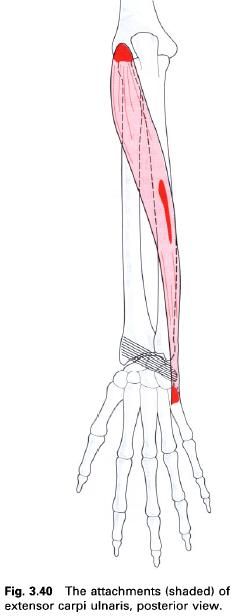
Extensor carpi ulnaris
Extensor carpi ulnaris arises from the lateral epicondyle of the humerus via the common extensor tendon and the adjacent fascia. There is also a
strong attachment, via a common aponeurosis
shared with flexor digitorum profundus
and flexor carpi ulnaris, from the
posterior border of the ulna. The
muscle forms a tendon near the wrist which passes below the extensor
retinaculum in its own synovial sheath and compartment. This is carried in a
groove next to the ulnar styloid process. The tendon attaches to a tubercle on
the medial side of the base of the fifth metacarpal.
Nerve
supply
Extensor carpi ulnaris is supplied by the
posterior interosseus branch of the radial nerve, root value C7, 8. The skin
over the muscles is supplied by roots C6, 7, 8.
Action
Working with extensor carpi radialis longus and
brevis produces extension of the wrist. The functional significance of this has
been described in the actions of the two former muscles. Working with flexor carpi ulnaris, extensor carpi
ulnaris produces adduction(ulnar deviation) at the wrist.
Palpation
The tendon of extensor carpi ulnaris can be
identified on the dorsum of the wrist, when the wrist is extended and adducted
against resistance. It lies on the lateral side of the ulnar styloid process.
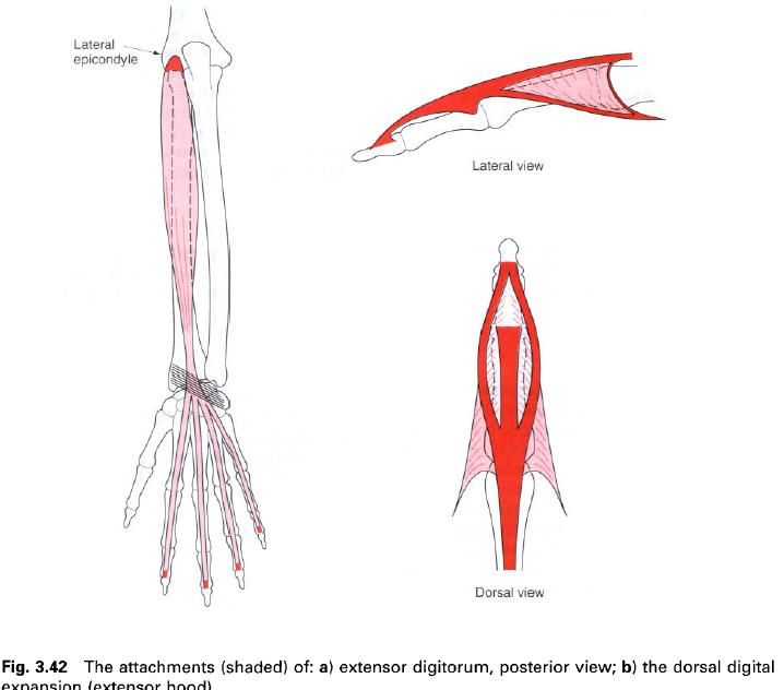
Extensor digitorum
Extensor digitorum is centrally placed within
the posterior compartment of the forearm. It arises from the lateral epicondyle of the humerus via the common extensor tendon, the covering fascia and the intermuscular
septa at its sides. In the lower part of the forearm the muscle forms four
tendons which pass deep to the extensor indicis. On the dorsum of the hand the tendons diverge towards the medial
four digits, being interconnected by obliquely placed fibrous bands. The
arrangement of these bands is variable, however, the tendons for the ring and
little fingers are attached usually until they pass just proximal to the
metacarpophalangeal joints. The tendon to the index finger may not be attached
to that of the middle finger.
The distal attachment of extensor digitorum is
complex in that each tendon helps to form an aponeurosis over the dorsum of the fingers. This is called the dorsal digital expansion or extensor
hood. In its simplest form it is best thought of as a movable triangular hood,
the base of which lies proximally over the metacarpophalangeal joint. From here
the sides of the hood wrap around the phalanx with the apex pointing distally.
The extensor hood forms the dorsal part of the capsule of the
metacarpophalangeal joint and extends forwards either side of the metacarpal
head to fuse with the deep transverse metacarpal ligament. As the extensor hood
approaches the proximal interphalangeal joint, it narrows and is reinforced on
either side by the interosseus and is reinforced on either side by the
interosseus and lumbrical muscles for
that finger. At the distal end of the proximal phalanx, the extensor hood
divides into three parts(figure b). The central part, which is directly
continuous with the extensor tendon, inserts into the base of the middle phalanx
on its dorsal aspect. The two collateral parts, which are continuous with the
tendons of the interossei and lumbricals,
reunite to insert into the base of
the distal phalanx on its dorsal
aspect. There may be an attachment to the base of the proximal phalanx but this
is unusual.
The way in which the extensor hood wraps around
the phalanges towards the palm facilitates the attachment of the lumbrical and
C7, 8. The skin over the muscle is supplied by roots C6, 7.
Action
Extensor indicis assists extensor digitorum in
its actions with respect to the joints of the index finger. It does, however, enable the index finger to be used
independently, and also aids extension of the wrist.
Palpation
The tendon of extensor indicis, lying medial to
that of extensor digitorum, can be palpated on the dorsum of the hand when the index finger is extended.
The muscle belly can be palpated by deep pressure over the lower part of the ulna during the same movement.
Extensor pollicis longus
Extensor pollicis longus lies deep to extensor
digitorum in the posterior compartment of the forearm. Here it arises from the
lateral part of the middle third of the
posterior surface of the ulna and the adjacent interosseus membrane above extensor
indicis. Above the wrist it forms a single tendon which passes in its own
synovial sheath deep to the extensor retinaculum, lying in a groove on the back
of the radius medial to the dorsal
tubercle. Winding around the dorsal tubercle, the tendon changes direction and
crosses the tendons of extensor carpi radialis longus and brevis and the radial
artery. As the tendon crosses the metacarpophalangeal joint it forms the dorsal
part of the joint capsule. It is joined in this region by slips from abductor
pollicis brevis laterally and adductor pollicis medially. The whole arrangement
forms a triangular expansion, not unlike that of extensor digitorum. The
expansion may also be joined by extensor pollicis brevis. The tendon of
extensor pollicis longus attaches to the dorsal
surface of the base of the distal
phalanx of the thumb. In its
course across the back of the hand, the tendon forms the medial boundary of a
region known as the anatomical “snuff-box”.
Nerve
supply
Extensor pollicis longus is supplied by the posterior interosseus branch of the radial nerve, root value C7, 8. The skin
over the muscle is supplied by roots C6 and 7.
Action
Extensor pollicis longus extends all of the
joints of the thumb, and assists in
extension and abduction of the wrist. Once this has been achieved, the
obliquity of the tendon will pull the first metacarpal into a laterally rotated
and abducted position. In full abduction or extension of the thumb, extensor pollicis longus can act
as an adductor. In addition, it may contribute slightly to supination, due to
its oblique course across the lower part of the forearm.
Palpation
The tendon of extensor pollicis longus is
easily identified with the thumb
extended as the medial border of the “anatomical snuff – box”. The tendon
frequently ruptures in conditions, such as rheumatoid arthritis or post Colles’
fracture. As a result, the patient is incapable of extending the
interphalangeal joint of the thumb.
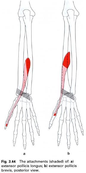
Extensor pollicis brevis
Extensor pollicis brevis lies on the lateral
side and is adjacent to extensor pollicis longus, and distal to abductor
pollicis longus to which it closely adheres. It arises from the middle part of the posterior surface of the radius
and adjacent interosseus membrane.
The tendon is formed above the wrist, and runs with that of abductor pollicis
longus deep to the extensor retinaculum in a common synovial sheath, in a
groove on the lateral surface of the radial styloid process. Below this point,
these two tendons form the lateral boundary of the “anatomical snuff – box”.
The tendon of extensor pollicis brevis partially replaces the dorsal part of
the capsule of the metacarpophalangeal joint of the thumb and inserts into the dorsal surface of the base of the
proximal phalanx. Occasionally the tendon may be prolonged, so that it runs
with and attaches to the tendon of extensor pollicis longus.
Nerve
supply
Extensor pollicis brevis is supplied by the posterior interosseus branch of the radial nerve, root value C7 and 8. The
skin over the muscle is supplied by roots C6 and 7.
Action
Extensor pollicis brevis extends both the
carpometacarpal and metacarpophalangeal joints of the thumb. It may also help in extending and abducting the wrist,
particularly against resistance.
Palpation
The tendons of extensor pollicis brevis and
abductor pollicis longus run together from the lower lateral aspect of the radius where they form the lateral
boundary of the “anatomical snuff – box”. Both tendons can be palpated when the
thumb is extended, that of abductor
pollicis longus being the most anteriorly placed.
Application
The synovial sheath of extensor pollicis brevis
and abductor pollicis longus frequently becomes inflamed in the region of the
radial styloid process(De Quervain’s syndrome). Techniques, such as transverse
frictions, ultrasound or injection can be usefully applied to this point.


0 коментара:
Постави коментар