Free Facebook Likes, Youtube Subscribers,  Twitter Followers

Ads 468x60px

Blogger Tricks

Blogger Themes

30. 6. 2012.

Adaptations to aerobic training – part I


Improvements in endurance that accompany regular(daily, every other day, etc.) aerobic training, such as running, cycling, or swimming, result from multiple adaptations to the training stimulus. Some adaptations occur within the muscles themselves, promoting more efficient transport and utilization of oxygen and fuel substrates. Still other important changes occur in the cardiovascular system, improving circulation to and within the muscles. Pulmonary adaptations occur to a lesser extent.
Aerobic training, or cardiorespiratory endurance training, improves central and peripheral blood flow and enhances the capacity of the muscle fibers to generate greater amounts of adenosine triphosphate(ATP).

Endurance: Muscular versus cardiorespiratory

Endurance is a term that refers to two separate but related concepts: muscular endurance and cardiorespiratory endurance. Each makes a unique contribution to athletic performance, and each differs in its importance to different athletes.
For sprinters, endurance is the quality that allows them to sustain a high speed over the full distance of, for example, a 100 or 200m race. This component of fitness is termed muscular endurance, the ability of a single muscle or muscle group to sustain high-intensity, repetitive, or static exercise. This type of endurance is also exemplified by the weightlifter doing multiple repetitions, the boxer, and the wrestler. The exercise or activity can be rhythmic and repetitive in nature, such as multiple repetitions of the bench press for the weightlifter and jabbing for the boxer. Or the activity can be more static, such as a sustained muscle action when a wrestler attempts to pin an opponent to the mat. In either case, the resulting fatique is confined to a specific muscle group, and the activity duration is usually no more than 1 to 2 min. Muscular endurance is highly related to muscular strength and to anaerobic power development.
While muscular endurance is specific to individual muscles or muscle groups, cardiorespiratory endurance relates to the entire body’s ability to sustain prolonged, dynamic exercise using large muscle groups. This type of endurance is used by the cyclist, distance runner, or endurance swimmer who completes long distances at a fairly fast pace. Cardiorespiratory endurance is related to the development of the cardiovascular and respiratory systems’ ability to maintain oxygen delivery to working muscles during prolonged bouts of exercise, as well as the muscles’ ability to utilize energy aerobically. This is why the terms cardiorespiratory endurance and aerobic endurance are sometimes used synonymously.

Evaluating cardiorespiratory endurance capacity

To study training effects on endurance, there needs to be an objective, repeatable means of measuring an individual’s cardiorespiratory endurance capacity. In that way, the exercise scientist, coach, or athlete can monitor improvements as physiological adaptations occur during the training program.

Maximal endurance capacity: VO2max or aerobic power

Most exercise scientists regard VO2max, sometimes called maximal aerobic power or maximal aerobic capacity, as the best objective laboratory measure of maximal cardiorespiratory endurance. VO2max is defined as the highest rate of oxygen consumption attainable during maximal or exhaustive exercise. VO2max is defined as the highest rate of oxygen consumption attainable during maximal or exhaustive exercise. VO2max as defined by the Fick equation is dictated by maximal cardiac output(delivery of oxygen and blood flow to working muscles) and the maximal(a-ṽ)O2 difference(the ability of the active muscles to extract and use the oxygen). As exercise intensity increases, oxygen consumption eventually either plateaus or decreases slightly, even with further increases in workload, indicating that a truly maximal VO2 has been achieved.
With endurance training, more oxygen can be delivered to, and consumed by, active muscles than in an untrained state. Previously untrained subjects demonstrate average increases in VO2max of 15% to 20% after a 20-week training program. These improvements allow individuals to perform endurance activities at a higher intensity, improving their performance potential. Figure below illustrates the increase of VO2max after 12 months of aerobic training in a previously untrained individual. In this example, VO2max increased by about 30%. Note that the VO2 “cost” of running at a certain submaximal intensity did not change but that higher running speeds could be attained after training.


Submaximal endurance capacity

In addition to increasing maximal endurance capacity, endurance training increases submaximal endurance capacity, which is much more difficult to evaluate. Steady-state submaximal heart rate at the same exercise intensity measured before and after training is one physiological variable that can be used to objectively quantify the effect of training. Additionally, exercise scientists have used performance measures to quantify submaximal endurance capacity. For example, one test used to determine submaximal endurance capacity is the average peak absolute power output a person can maintain over a fixed period of time on a cycle ergometer. For running, the average peak speed of velocity a person can maintain during a fixed period of time would be similar type of test. Generally, these tests will last 30 min to an hour.
Submaximal endurance capacity is more closely related to actual competitive endurance performance than VO2max, and is likely determined by both the person’s VO2max and the threshold for his or her onset of blood lactic acid accumulation(OBLA) – that point at which lactate begins to appear at a disproportionate rate in the blood. With endurance training, submaximal endurance capacity increases.

Cardiovascular adaptations to training

Numerous cardiovascular adaptations occur in response to exercise training, including changes in the following cardiovascular variables:
  • Heart size
  • Stroke volume
  • Heart rate
  • Cardiac output
  • Blood flow
  • Blood pressure
  • Blood volume.

To understand completely, it is very important to review how these components relate to oxygen transport.

Oxygen transport system

Cardiorespiratory endurance is related to the cardiovascular and respiratory systems’  ability to deliver sufficient oxygen to meet the needs of metabolically active tissues.
The ability of the cardiovascular and respiratory systems to deliver oxygen to active tissues is defined by Fick equation. The Fick equation states that systemic oxygen consumption is determined by both the delivery of oxygen(cardiac output) via blood flow and the amount of oxygen extracted by the tissues, the (a-ṽ)O2 difference. The product of cardiac output and the (a-ṽ)O2 difference determines the rate of which oxygen is being consumed:

VO2 = stroke volume x heart rate x (a-ṽ)O2 diff.

The oxygen demand of exercising muscles increases with increasing exercise intensity. Aerobic endurance depends on the cardiorespiratory system’s ability to deliver sufficient oxygen to these active tissues to meet their heightened demands for oxygen for oxidative metabolism. As maximal levels of exercise are achieved, heart size, blood flow, blood pressure, and blood volume can all potentially limit the maximal ability to transport oxygen. Endurance training elicits numerous changes in these components of the oxygen transport system that enable it to function more effectively.

Heart size

As an adaptation to the increased work demand, heart mass and volume increase with training. Cardiac muscle, like skeletal muscle, undergoes morphological adaptations as a result of chronic endurance training. At one time, cardiac hypertrophy induced by exercise – “athlete’s heart”, as it was called – caused some concern because experts incorrectly believed that enlargement of the heart always reflected a pathological state, as sometimes occurs with severe hypertension. Training-induced cardiac hypertrophy is now recognized as a normal adaptation to chronic endurance training.
The left ventricle, does the most work and thus undergoes the greatest adaptation in response to endurance training. The extent and location of heart size adaptations depend on the type of exercise training performed. For example, during resistance training, the left ventricle must contract against increased afterload from the systemic circulation. It was postulated to overcome this high afterload, the heart muscle compensated by increasing left ventricular wall thickness, thereby increasing its contractility. Blood pressure during resistance training can exceed 480/350 mmHg. This presents a considerable resistance that must be overcome by the left ventricle. Thus, the increase in its muscle mass is in direct response to repeated exposure to the increased afterload with resistance training.
With endurance training, left ventricular chamber size increases. This allows for increased left ventricular filling and consequently an increase in stroke volume. The increase in left ventricular dimensions is largely attributable to a training-induced increase in plasma volume that increases left ventricular end-diastolic volume(increased preload). In concert with this, a decrease in heart rate at rest caused by increased parasympathetic tone, and during exercise at the same rate of work, allows a longer diastolic filling period. The increases in plasma volume and diastolic filling time increase left ventricular chamber size at the end of diastole.
It was originally hypothesized that this increase in left ventricular dimensions was the only change in the left ventricle caused by endurance training. Additional research has revealed that myocardial wall thickness also increases with endurance training, rather than just with resistance training. Using magnetic resonance imaging, Milliken and colleagues found that highly trained endurance athletes(competitive cross-country skiers, endurance cyclists, and long-distance runners) had greater left ventricular masses than did non-endurance-trained control subjects. Left ventricular mass was highly correlated with VO2max or aerobic power.
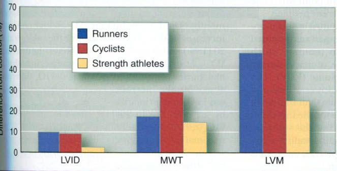
Fagard conducted the most extensive review of the existing research literature in 1996, focusing on long-distance runners(135 athletes and 173 controls), cyclists(69 athletes and 65 controls), and strength athletes(178 athletes, including weight- and powerlifters, bodybuilders, wrestlers, throwers, and bobsledders, and 105 controls). For each group, the athletes were matched by age and body size with a group of sedentary control subjects. For each group of runners, cyclists, and strength athletes, the internal diameter of the left ventricle(LVID, an index of chamber size) and the total left ventricular mass(LVM) were greater in the athletes compared with their age- and sized-matched controls(figure below). Thus, data from this large cross-sectional study support the hypothesis that both left ventricular chamber size and wall thickness increase with endurance training.


Most studies of the heart size changes with training have been cross-sectional, comparing trained individuals with sedentary, untrained individuals. We can learn much from cross-sectional studies, but they do not provide us with the same information that we could get from studying a group of untrained people who train for months or years, focusing on their changes from the untrained to the trained state. Certainly a portion of the differences that we see in figure above us can be attributed to genetics, not training. However, a number of studies have followed individuals from an untrained state to a trained state to an untrained state. These studies have reported increases in heart size with training and decreases with detraining. So, training does bring about changes, but they are likely not as large as the differences we see in the figure above us.

Stroke volume

As a result of endurance training, stroke volume increases. Stroke volume at rest is substantially higher after an endurance training program than it is before training. This endurance training-induced increase is also seen at a 
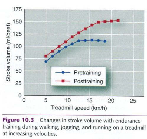
given submaximal exercise intensity and at maximal exercise. This increase is illustrated in figure below, which shows the changes in stroke volume of a subject who exercised at increasing intensities up to maximal intensity before and after a six-month aerobic endurance training program. Typical values for stroke volume at rest and during maximal exercise in untrained, trained, and highly trained athletes are listed in the table below. The wide variation in stroke volume values for any given cell within this table is largely attributable to differences in body size. Absolute stroke volume at rest and during exercise is not merely a function of a person’s state of training but also reflects differences in body size. Larger people typically have larger hearts and a greater blood volume, and thus higher stroke volumes – an important point when one is comparing stroke volumes of different people.


Stroke volumes(SV) for different states of training
Subjects
SVrest(ml/beat)
SVmax(ml/beat)
Untrained
50-70
80-110
Trained
70-90
110-150
Highly trained
90-110
150- >220

After aerobic training, the left ventricle fills more completely during diastole than it does in an untrained state. Plasma volume expands with training, which allows for more blood to enter the ventricle during diastole, increasing end-diastolic volume(EDV). The heart rate of a trained heart is also lower at rest and at the same absolute exercise intensity than that of an untrained heart, allowing more time for the increased diastolic filling. More blood entering the ventricle increases the stretch on the ventricular walls; by the Frank-Starling mechanism, this results in an increase in force of contraction.
The thickness of the posterior and septal walls of the left ventricle also increases slightly with endurance training. Increased ventricular muscle mass results in increased contractile force, in turn causing end-systolic volume to decrease. More blood is forced out of the heart, leaving less blood in the left ventricle after systole.
The decrease in end-systolic volume is augmented by the decrease in peripheral resistance that occurs with training. Increased contractility resulting from an increase in left ventricular thickness and greater diastolic filling(Frank-Starling mechanism), coupled with the reduction in systemic peripheral resistance, increases the ejection fraction[equal to (EDV-ESV)/EDV] in the trained heart. More blood enters the left ventricle, and a greater percentage of what enters is forced out with each concentration, resulting in an increase in stroke volume.
Adaptatioins in stroke volume during endurance training are illustrated by a study in which older men endurance trained for one year. Their cardiovascular function was evaluated before and after training. The subjects performed running, treadmill, and cycle ergometer exercise for 1h each day, four days per week. They exercised at intensities of 60% to 80% pf VO2max, with brief bouts of exercise exceeding 90% of VO2max. End-diastolic volume increased at rest and throughout submaximal exercise. The ejection fraction increased, which was associated with a decreased end-systolic volume, suggesting increased contractility of the left ventricle. VO2max increased by 23%, indicating a substantial improvement in endurance.
It is clear that central stroke volume adaptations occur with endurance training, but there are also peripheral adaptations that contribute to the increase in VO2max, at least in middle-aged exercisers. This was demonstrated in a unique longitudinal study involving both exercise training and a bed rest deconditioning model. Five 20-year-old men were tested(baseline values), placed on bed rest for 20 days(deconditioning), and then trained for 60 days, starting immediately at the conclusion of bed rest. These same five men were restudied 30 years later at the age of 50; they were tested at baseline in a relatively sedentary state and after six months of endurance training. The average percentage increases in VO2max were similar for the subjects at age 20(18%) and at age 50(14%). However, the increase in VO2max at age 20 was explained by increases in both maximal cardiac output and maximal(a-ṽ)O2 difference; at age 50, the increase was explained primarily by an increase in(a-ṽ)O2 difference, while maximal cardiac output was unchanged. Maximal stroke volume was increased after training at both age 20 and age 50 but to a lesser degree at age 50(+16ml/beat at age 20 vs. +8ml/beat at age 50).

29. 6. 2012.

Resistance training for special populations


Until the 1970s, resistance training was widely regarded as appropriate only for young, healthy, male athletes. This narrow concept led many people to overlook the benefits of resistance training when planning their own activities. First sex and age differences will be considered, and then this form of training for the athletes, regardless of sex, age, or sport.

Sex and age differences

In recent years, considerable interest has focused on training for women, children, and people who are elderly. As mentioned earlier, the widespread use of resistance training by women, either for sport or for health-related benefits, is rather recent. Substantial knowledge has developed since the early 1970s revealing that women and men have the same ability to develop strength but that, on average, women may not be able to achieve peak values as high as those attained by men. This difference in strength is attributable primarily to muscle size differences related to sex differencies in anabolic hormones. Resistance training techniques developed for and applied to men’s training seem equally appropriate for women’s training.
The wisdom of resistance training for children and adolescents has long been debated. The potential for injury, particularly growth plate injuries from the use of free weights, has caused much concern. Many people once believed that children would not benefit from resistance training, based on the assumption that the hormonal changes associated with puberty are necessary for gaining muscle strength and mass. We now know that children and adolescents can train safely with minimal risk of injury if appropriate safeguards are followed. Furthermore, they can indeed gain both muscular strength and muscle mass.
Interest in resistance training procedures for elderly people has also increased. A substantial loss of fat-free body mass accompanies aging. This loss of reflects mainly the loss of muscle mass, largely because most people become less active as they age. When a muscle isn’t used regularly, it loses function, with predictable atrophy and loss of strength.  
Can resistance training in elderly people reverse this process? People who are elderly can indeed gain strength and muscle mass in response to resistance training. This fact has important implications for both their health and the quality of their lives. With maintained or improved strength, they are less likely to fall. This is a significant benefit because falls are a major source of injury and debilitation for elderly people and often lead to death.

Resistance training for sport

Gaining strength, power, or muscular endurance simply for the sake of being stronger, being more powerful, or having greater muscular endurance is of relatively little importance to athletes unless it also improves their athletic performance. Resistance training by field-event athletes and competitive weightlifters makes intuitive sense. The need for resistance training by the gymnast, distance runner, baseball player, high jumper, or ballet dancer is less obvious.
We do not have extensive research to document the specific benefits of resistance training for every sport of for every event within a sport. But clearly each has basic strength, power, and muscular endurance requirements that must be met to achieve optimal performance. Training beyond these requirements may be unnecessary.
Training is costly in terms of time, and athletes can’t afford to waste time on activities that won’t result in better athletic performances. Thus, some performance measurement is imperative to evaluate any resistance training program’s efficacy. To resistance train solely to become stronger, with no associated improvement in performance, is of questionable value. However, it should also be recognized that resistance training can reduce the risk of injury for most sports, because fatiqued individuals are at an increased risk of injury.

Muscle soreness


Muscle soreness generally results from exhaustive or very high intensity exercise. This is particularly true when people perform a specific exercise for the first time. While muscle soreness can be felt at any time, there is generally a period of mild muscle soreness that can be felt during and immediately after exercise, and then a more intense soreness felt a day or two later.

Acute muscle soreness

Pain felt during and immediately after exercise can result from accumulation of the end products of exercise, such as H+, and from tissue edema, mentioned earlier, which is caused by fluid shifting from the blood plasma into the tissues. Edema is the cause of the pumped-up sensation that people feel after heavy endurance or strength training. The pain and soreness usually disappear within a few minutes to several hours after the exercise. Thus, this soreness is often referred to as acute muscle soreness.

Delayed-onset muscle soreness and injury

Muscle soreness felt a day or two after a heavy bout of exercise is not totally understood, yet researchers are continuing to give us greater insight into this phenomenon. Because the pain does not occur immediately, it is reffered to as delayed-onset muscle soreness(DOMS). Here will be discussed theories that attempt to explain this form of muscle soreness.
Almost all current theories acknowledge that eccentric action is the primary initiator of DOMS. This was clearly demonstrated in a study of the relationship of muscle soreness to eccentric, concentric, and static actions. This study showed that a group who trained solely with eccentric actions experienced extreme muscle soreness, whereas the static- and concentric- action groups experienced little soreness. This idea was further explored in studies in which subjects ran on a treadmill for 45 min on two separate days, one day on a level grade and the other day on a 10% downhill grade.
No muscle soreness was associated with the level running. But the downhill running, which required extensive eccentric action, resulted in considerable soreness within 24 to 48h, even though blood lactate levels, previously thought to cause muscle soreness, were much higher with level running.

Structural damage

The presence of increased concentrations of several specific muscle enzymes in blood after intense exercise suggests that some structural damage may occur in the muscle membranes. These enzyme levels increase from 2 to 10 times their normal levels following bouts of heavy training. Recent studies support the idea that these changes might indicate various degrees of muscle tissue breakdown. Examination of tissue from the leg muscles of marathon runners has revealed remarkable damage to the muscle fibers after both training and marathon competition. The onset and timing of these muscle changes parallel the degree of muscle soreness experienced by the runners.
Although the effects of muscle damage on performance are not fully understood, experts generally agree that this damage is responsible in part for the localized muscle pain, tenderness, and swelling associated with DOMS. However, blood enzyme levels might increase and muscle fibers might be damaged frequently during daily exercise that produces no muscle soreness. Also, remember that muscle damage appears to be a precipitating factor for muscle hypertrophy.

Inflammatory reaction

White blood cells serve as a defense against foreign materials that enter the body and against conditions that threated the normal function of tissues. The white blood cell count tends to increase following activities that induce muscle soreness. This observation led some investigators to suggest that soreness results from inflammatory reactions in the muscle. But the link between these reactions and muscle soreness has been difficult to establish.
Researchers have tried to use drugs to block the inflammatory reaction, but these efforts have been unsuccessful in reducing the amount of muscle soreness or the degree of inflammation. Because both effects remain, conclusions about the role of inflammation in muscle soreness cannot be drawn from this research. More recent studies, however, are beginning to establish a link between muscle soreness and inflammation. For example, it is now recognized that substances released from injured muscle can act as attractants, initiating the inflammatory process. Mononucleated cells in muscle are activated by the injury, providing the chemical signal to circulating inflammatory cells. Neutrophils(a type of white blood cell) invade the injury site and release cytokines(immunoregulatory substances), which then attract and activate additional inflammatory cells. Neutrophils possibly also release oxygen free radicals that can damage cell membranes. Macrophages(another type of cell of the immune system) then invade the damaged muscle fibers, removing debris through a process known as phagocytosis. Last, a second phase of macrophage invasion occurs, which is associated with muscle regeneration.

Sequence of events in DOMS

In 1984, Armstrong reviewed possible mechanisms for exercise-induced DOMS. He concluded that DOMS is associated with:
  • Elevations in plasma enzymes,
  • Myoglobinemia(presence of myoglobin in the blood), and
  • Abnormal muscle histology and ultrastructure.

He developed a model of DOMS that proposed the following sequence of events:
1.            High tension in the contractile-elastic system of muscle results in structural damage to the muscle and its cell membrane.
2.            The cell membrane damage disturbs calcium homeostasis in the injured fiber, resulting in necrosis(cell death) that peaks about 48h after exercise.
3.            The products of macrophage activity and intracellular contents(such as histamine, kinins, and K+) accumulate outside the cells. These substances then stimulate the free nerve endings in the muscle. This process appears to be accentuated in eccentric exercise, in which large forces are distributed over relatively small cross-sectional areas of the muscle.

Recent comprehensive reviews have provided much greater insight into the cause of muscle soreness. We now are confident that muscle soreness results from injury or damage to the muscle itself, generally the muscle fiber and possibly the plasmalemma. This damage sets up a chain of events that includes the release of intracellular proteins and an increase in muscle protein turnover. The damage and repair process involves calcium ions, lysosomes, connective tissue, free radicals, energy sources, inflammatory reactions, and intracellular and myofibrillar proteins. But the precise cause of skeletal muscle damage and the mechanisms of repair are not well understood. Some evidence suggests that this process is an important step in muscle hypertrophy.
Up to this point, our discussion of DOMS has focused on muscle injury. Edema, or the accumulation of fluids in the muscular compartment, also can lead to DOMS. This edema is likely the result of muscle injury but could occur independently of muscle injury. An accumulation or interstitial or intracellular fluid increases the tissue fluid pressure within the muscle compartment, which in turn activates pain receptors within the muscle.

Delayed-onset muscle soreness and performance

With DOMS comes a reduction in the force-generating capacity of the affected muscles. Whether the DOMS is the result of injury to the muscle or edema independent of muscle injury, the affected muscles are not able to exert as much force when the person is asked to apply maximal force, such as in the performance of a 1RM strength test. Maximal force-generating capacity gradually returns over days or weeks. It has been proposed that the loss in strength is the result of three factors:
  1. The physical disruption of the muscle ,
  2. Failure within the excitation – contraction coupling process,
  3. Loss of contractile protein.

Failure in excitation – contraction coupling appears to be the most important, particularly during the first five days.


Muscle glycogen resynthesis also is impaired when a muscle is damaged. Resynthesis is generally normal for the first 6 to 12h after exercise, but it slows or stops completely as the muscle undergoes repair, thus limiting the fuel-storage capacity of the injured muscle. Figure below illustrates the time sequence of the various factors associated with intense eccentric exercise, including pain, edema, plasma creatine kinase( a plasma enzyme marker of muscle fiber damage), glycogen depletion, ultrastructural damage in the muscle, and muscular weakness.


Reducing the negative effects of DOMS

Reducing the negative effects of DOMS is important for maximizing training gains. The eccentric component of muscle action could be minimized during early training, but this is not possible for athletes in most sports. An alternative approach is to start training at a very low intensity and progress slowly through the first few weeks. Yet another approach is to initiate the training program with a high-intensity, exhaustive training bout. Muscle soreness would be great for the first few days, but evidence suggests that subsequent training bouts would cause considerably less muscle soreness. Because the factors associated with DOMS are also potentially important in stimulating muscle hypertrophy, DOMS is most likely necessary to maximize the training response.

Muscle atrophy and decreased strength with inactivity


When a normally active or highly trained person reduces his or her level of activity or ceases training altogether, major changes occur in both muscle structure and function. This is illustrated in the picture below by the results of two studies: studies in which entire limbs have been immobilized and studies in which highly trained people stop training.


Immobilization

When a trained muscle suddenly becomes inactive through immobilization, major changes are initiated within that muscle in a matter of hours. During the first 6h of immobilization, the rate of protein synthesis starts to decrease. This decrease likely initiates the start of muscular atrophy, which is the wasting away or decrease in the size of muscle tissue. Atrophy results from lack of muscle use and the consequent loss of muscle protein that accompanies the inactivity. Strength decreases are most dramatic during the first week of immobilization, averaging 3% to 4% per day. This is associated with the atrophy but also with decreased neuromuscular activity of the immobilized muscle.
Immobilization appears to affect both type I and type II fibers. From various studies, researchers have observed disintegrated myofibrils, streaming Z-disks(discontinuity of Z-disks and fusion of the myofibrils), and mitochondrial damage. When muscle atrophies, the cross-sectional fiber area decreases. Several studies have shown the effect to be greater in type I fibers, including a decrease in the percentage of type I fibers, thereby increasing the percentage of type II fibers.
Muscles can and often do recover from immobilization when activity is resumed. The recovery period is substantially longer than the period of immobilization but shorter than the original training period.

Cessation of training

Similarly, significant muscle alterations can occur when people stop training. In one study, women resistance trained for 20 weeks and then stopped training for 30 to 32 weeks. Finally they retrained for six weeks. The training program focused on the lower extremity, using a full squat, leg press, and leg extension. Strength increases were dramatic, as seen in the figure below. Compare the women’s strength after their initial training period(post-20) with their strength after detraining(pre-6). This represents the strength loss they experienced with cessation of training.


During the two training periods, increases in strength were accompanied by increases in the cross-sectional area of all fiber types and a decrease in the percentage of type IIx fibers. Detraining had relatively little effect on fiber cross-sectional area, although the type II fiber areas tended to decrease.


To prevent losses in the strength gained through resistance training, basic maintenance programs must be established once the desired goals for strength development have been achieved. Maintenance programs are designed to provide sufficient stress to the muscles to maintain existing levels of strength while allowing a reduction in intensity, duration, or frequency of training.
In one study, men and women resistance trained with knee extensions for either 10 or 18 weeks and then spent an additional 12 weeks with either no training or reduced training. Knee extension strength increased 21.4% following the training period. Subjects who then stopped training lost 68% of their strength gains during the weeks they didn’t train. But subjects who reduced their training(from three days per week to two, or from two to one) did not lose strength. Thus, it appears that strength can be maintained for at least up to 12 weeks with reduced training frequency.

Fiber type alterations

Can muscle fibers change from one type to another through resistance training? The earliest research included that neither speed(anaerobic) nor endurance(aerobic) training could after the basic fiber type, specifically from type I to type II, or from type II to type I. These early studies did show, however, that fibers begin to take on certain characteristics of the opposite fiber type if the training is of the opposite kind(e.g. type II fibers might become more oxidative with aerobic training).
Research with animals has shown that fiber type conversion is indeed possible under conditions of cross-innervation, in which a type II motor unit is artificially innervated by a type I motor neuron or vice versa. Also, chronic, low-frequency nerve stimulation transforms type II motor units into type I motor units within a matter of weeks. Muscle fiber types in rats have changed in response to 15 weeks of high-intensity treadmill training, resulting in an increase in type I and type IIa fibers from type IIx to type IIa and from type IIa to type I was confirmed by several different histochemical techniques.
Staron and coworkers found evidence of fiber type transformation in women as a result of heavy resistance training. Substantial increases in static strength and in the cross-sectional area of all fiber types were noted following a 20-week heavy resistance training program for the lower extremity. The mean percentage of type IIx fibers decreased significantly, but the mean percentage of type IIa fibers increased. The transition of type IIx fibers to type IIa fibers with resistance training has been consistently reported in a number of subsequent studies. More recent studies have shown that a combination of high-intensity resistance training training and short-interval speed work can lead to a conversion of type I and type IIa fibers.

Integration of neural activation and fiber hypertrophy


Research on resistance training adaptations indicates that early increases in voluntary strength, or maximal force production, are associated primarily with neural adaptations resulting in increased voluntary activation of muscle. This was clearly demonstrated in a study of both men and women who participated in an eight-week, high-intensity resistance training program, training twice per week. Muscle biopsies were obtained at the beginning of the study and every two weeks during the training period. Strength, measured according to the 1RM, increased substantially over the eight weeks of training, with the greatest gains coming after the second week. Muscle biopsies, however, revealed only small, statistically insignificant increases in muscle fiber cross-sectional area by the end of the eight weeks of training. Thus, the strength gains were largely the result of increased neural activation.
Long-term increases in strength generally are associated with hypertrophy of trained muscle. It takes time to build protein through a decrease in protein degradation, an increase in protein synthesis, or both. Notable exceptions to this generalization have been found. A six-month study of strength-trained athletes showed that neural activation explained most of the strength gains during the most intensive training months and that hypertrophy was not a major factor. It appears that neural factors make their greatest contribution during the first 8 to 10 weeks of training. Hypertrophy contributes little during the initial weeks of training but progressively increases its contribution, becoming the major contributor after 10 weeks of training.

Fiber hyperplasia


Research on animal suggests that hyperplasia may also be a factor in the hypertrophy of whole muscles. Studies on cats provide fairly clear evidence that fiber splitting occurs with extremely heavy weight training. Cats were trained to move a heavy weight with a forepaw to get their food.


They learned to generate considerable force. With this intense strength training, selected muscle fibers appeared to actually split in half, and each half then increased to the size of the parent fiber. This is seen in the cross-sectional cuts through the muscle fibers shown in figure below.


Subsequent studies, however, demonstrated that hypertrophy of selected muscles in chickens, rats, and mice that resulted from chronic exercise overload was attributable solely to hypertrophy of existing fibers, not hyperplasia. In these studies, each fiber in the whole muscle was actually counted. These direct fiber counts revealed no change in fiber number.
This finding led the scientists who conducted the initial cat experiments to conduct an additional resistance training study with cats. This time they used actual fiber counts to determine if total muscle hypertrophy resulted from hyperplasia or fiber hypertrophy. Following a resistance training program of 101 weeks, the cats were able to perform one-leg lifts of an average of 57% of their body weight, resulting in an 11% increase in muscle weight. Most important, the researchers found a 9% increase in the total number of muscle fibers, confirming that muscle fiber hyperplasia did occur.
The difference in results between the cat studies and those with rats and mice most likely is attributable to differences in the manner in which the animals were trained. The cats were trained with a pure form of resistance training: high resistance and low repetitions. The other animals were trained with more endurance type activity: low resistance and high repetitions.
One additional animal model has been used to stimulate muscle hypertrophy associated with hyperplasia. Scientists have placed the anterior latissimus dorsi muscle of chickens in a state of chronic stretch by attaching weights to it, with one other wing serving as the normal control condition. In many of the studies that have used this model, the chronic stretch has resulted in substantial hypertrophy and hyperplasia.
Researchers are still uncertain about the roles played by hyperplasia and individual fiber hypertrophy in increasing human muscle size with resistance training. Most evidence indicates that individual fiber hypertrophy accounts for most whole-muscle hypertrophy. However, results from selected studies indicate that hyperplasia is possible in humans.
In several studies of bodybuilders, swimmers, and kayakers, substantial muscle hypertrophy has been observed in trained muscles, but in the absence of individual fiber hypertrophy when compared to values in untrained control subjects. This suggests that there is a greater number of muscle fibers in the trained muscles than in the corresponding musclesof untrained control subjects. However, other studies have shown individual fiber hypertrophy in highly trained athletes compared to untrained controls.
In a study of seven previously healthy young men who had suffered sudden accidental death, the investigators compared cross sections of autopsied right and left tibialis anterior muscles(lower leg). Right-hand dominance is known to lead to greater hypertrophy of the left leg. In fact, the average cross-sectional area of the left muscle was 7.5% larger. This was associated with a 10% greater number of fibers in the left muscle. There was no difference in fiber size.
The differences among these studies mighte be explained by the nature of the training load or stimulus. Training at high intensities or high resistances is thought to cause greater fiber hypertrophy, particularly of the type II(fast-twitch) fibers, than training at lower intensities or resistances.
Only one longitudinal study demonstrated the possibility of hyperplasia in men who had previous recreational resistance training experience. Following 12 weeks of intensified resistance training, the muscle fiber number in the biceps brachii of several of the 12 subjects appeared to increase significantly. It appears from this study that hyperplasia can occur in humans, but possibly only in certain subjects or under certain training conditions.
From the preceding information, it appears that fiber hyperplasia can occur in animals and possibly in humans. How are these cells formed? As shown in the previous picture, it is postulated that individual muscle fibers have capacity to divide and split into two daughter cells, each of which can then develop into a functional muscle fiber. It has more recently been established that satellite cells, which are the myogenic stem cells involved in skeletal muscle regeneration, are likely involved in the generation of new muscle fibers. These cells are typically activated by muscle stretching and injury, and, muscle injury results from intense training, particularly eccentric-action training. Muscle injury can lead to a cascade of responses, in which satellite cells become activated and proliferate, migrate to the damaged region, and fuse to existing myofibers or combine and fuse to produce new myofibers.


28. 6. 2012.

Muscle hypertrophy


How does a muscle size increase? Two types of hypertrophy can occur: transient and chronic. Transient hypertrophy is the increased muscle size that develops during and immediately following a single exercise bout. This results mainly from fluid accumulation(edema) in the interstitial and intracellular spaces of the muscle that comes from the blood plasma. Transient hypertrophy, as its name implies, lasts only for a short time. The fluid returns to the blood within hours after exercise.
Chronic hypertrophy refers to the increase in muscle size that occurs with long-term resistance training. This reflects actual structural changes in the muscle that can result from an increase in the size of existing individual muscle fibers(fiber hypertrophy), in the number of muscle fibers(fiber hyperplasia), or both. Controversy surrounds the theories that attempt to explain the underlying cause of this phenomenon. Of importance, however, is the finding that the eccentric component of training is important in maximizing increases in muscle fiber cross-sectional area. A number of studies have shown greater hypertrophy and strength resulting solely from eccentric contraction training as compared to concentric contraction or combined eccentric and concentric contraction training. Further, higher-velocity eccentric training appears to result in greater hypertrophy and strength gains than slower-velocity training. These greater increases appear to be related to disruptions in the sarcomere Z-lines. This disruption had originally been labeled as muscle damage, but is now thought to represent fiber protein remodeling. Thus, training with only concentric actions could limit muscle hypertrophy and increases in muscle strength.

Fiber hypertrophy

Early research suggested that the number of muscle fibers in each of a person’s muscles is established by birth or shortly thereafter and that this number remains fixed throughout life. If this were true, then wholemuscle hypertrophy could result only from individual muscle fiber hypertrophy. This could be explained by:

Intense resistance training can significantly increase the cross-sectional area of muscle fibers. For example, take a man who did not train two years, and returns to normal program and trains six months. His fiber size will be significantly bigger, and more cross-bridges for force production will appear in his legs. Assume is that hypertrophy will be caused by increased numbers of myofibrils and actin and myosin filaments, but this will not be the cause every single time.
Individual muscle fiber hypertrophy from resistance training appears to result from a net increase in muscle protein synthesis. The muscle’s protein content is in continual state of flux. Protein is always being synthetized and degraded. But the rates of these processes vary with the demands placed on the body. During exercise, protein synthesis decreases, while protein degradation apparently increases. This pattern reverses during the postexercise recovery period, even to the point of a net synthesis of protein. The provision of a carbohydrate and protein supplement immediately after a training bout can create a more positive nitrogen balance, facilitating protein synthesis.
The hormone testosterone is thought to be at least partly responsible for these changes, because one of its primary functions is the promotion of muscle growth. For example, males experience a significantly greater increase in muscle growth starting at puberty, which is largely due to a 10-fold increase in testosterone production. Testosterone is a steroidal hormone with major anabolic functions. It has been well established that massive doses of anabolic steroids coupled with resistance training markedly increase muscle mass and strength.

Mechanisms of gains of muscle strength


For many years, strength gains were assumed to result directly from increases in muscle size(hypertrophy). This assumption was logical because most who strength trained regularly were men, and they often developed large, bulky muscles. Also, muscles associated with a limb immobilized in a cast for weeks or months start to decrease in size(atrophy) and lose strength almost immediately. Gains in muscle size are generally paralleled by gains in strength, and loses in muscle size correlate highly with losses in strength. Thus, it is tempting to conclude that a direct cause-and-effect relationship exists between muscle size and muscle strength. While there is a relationship between size and strength, muscle strength involves far more than mere muscle size.
Numerous media reports indicate that people can perform superhuman feats of strength during great psychological stress. Straitjackets were designed specifically to control patients in mental hospitals who suddenly go berserk and are impossible to restrain. Even the world of sport boasts isolated examples of superhuman athletic performances, such as Bob Beamon’s long jump of 29ft(2 ½ inches – 8,90m) at the 1968 Olympic Games – a jump that exceeded the previous world record by nearly 2ft(0.6m)! World records are usually broken by inches or centimeters or, more often, mere fractions of inches or centimeters. Beamon’s record stood unbroken till 1991!
Women experience similar, or even greater, percentage increases in strength compared with men who participate in the same training program, but women generally do not experience as much hypertrophy. Similar findings have been reported in children.
This does not mean that muscle size is unimportant in the ultimate strength potential of the muscle. Size is extremely important, as revealed by the existing men’s and women’s world records for competitive weight-lifting.  As weight classification increases(implying increased muscle mass), so does the record for the total weight lifted. However, examples of superhuman strength and studies on women and children indicate that the mechanisms associated with strength gains are very complex and are not completely understood at this time. Obviously, increased muscle size is important, but there is increasing evidence that the neural control of the trained muscle is also altered, allowing a greater force production from the muscle.

Neural control of strength gains

An important neural component explains at least some of the strength gains that result from resistance training. Enoka has made a convincing argument that strength gains can be achieved without structural changes in muscle but not without neural adaptations. Thus, strength is not solely a property of the muscle. Rather, it is a property of the motor system. Motor unit recruitment, stimulation frequency, and other neural factors are also quite important to strength gains. They may well explain most, if not all, strength gains that occur in the absence of hypertrophy, as well as episodic superhuman feats of strength.

Synchronization and recruitment of additional motor units

Motor units are generally recruited asynchronously; they are not all called on at the same instant. They are controlled by a number of different neurons that can transmit either excitatory or inhibitory impulses. Whether the muscle fibers contract or stay relaxed depends on the summation of the many impulses received by the given motor unit at any one time. The motor unit is activated and its muscle fibers contract only when the incoming excitatory impulses exceed the inhibitory impulses and the threshold is met.
Strength gains may result from changes in the connections between motor neurons located in the spinal cord, allowing motor units to act more synchronously, facilitating contraction, and increasing the muscle’s ability to generate force. There is good evidence to support increased motor unit synchronization with resistance training; but there is still controversy as to whether synchronization of motor unit activation produces a more forceful contraction. It is clear, however, that synchronization does improve the rate of force development and the capability to exert steady forces.
An alternate possibility is simply that more motor units are recruited to perform the given task, independent of whether these motor units act in unison. Such improvement in recruitment patterns could result from an increase in neural drive to the alpha-motor neurons during maximal contraction. This increase in neural drive could also increase the frequency of discharge(rate coding) of the motor units. It is also possible that the inhibitory impulses are reduced, allowing more motor untis to be activated, or to be activated at a higher frequency.

Increased rate coding of motor units

The increase in neural drive of alpha-motor neurons could also increase the frequency of discharge, or rate coding, of their motor units. Recall from previous threads that as the frequency of stimulation of a given motor unit is increased, the muscle eventually reaches a state of tetanus, producing the absolute peak force or tension of the muscle fiber or motor unit. There is limited evidence that rate coding is increased with resistance training. Rapid movement or ballistic-type training appears to be particularly effective in stimulating increases in rate coding.

Autogenic inhibition

Inhibitory mechanisms in the neuromuscular system, such as the Golgi tendon organs, might be necessary to prevent the muscles from exerting more force than the bones and connective tissues can tolerate. This control is referred to as autogenic inhibition. During superhuman feats of strength, major damage often occurs to these structures, suggesting that the protective inhibitory mechanisms are overridden.
When the tension on a muscle’s tendons and internal connective tissue structures exceeds the threshold of the embedded Golgi tendon organs, motor neurons to that muscle are inhibited; that is, autogenic inhibition occurs. Both the reticular formation in the brain stem and the cerebral cortex function to initiate and propagate inhibitory impulses.
Training can gradually reduce or counteract these inhibitory impulses, allowing the muscle to reach greater levels of strength. Thus, strength gains may be achieved by reduced neurological inhibition. This theory is attractive because it can at least partially explain superhuman feats of strength and strength gains in the absence of hypertrophy.

Other neural factors

In addition to increasing motor unit recruitment or decreasing neurological inhibition, other neural factors can contribute to strength gains with resistance training. One of these is referred to as coactivation of agonist and antagonist muscles(the agonist muscles are the primary movers, and the antagonist muscles act to impede agonists). If we use forearm flexor concentric contraction as an example, the biceps is the primary agonist and the triceps is the antagonist. If both were contracting with equal force development, no movement would occur. Thus, to maximize the force generated by an agonist, it is necessary to minimize the amount of coactivation. Reduction in coactivation could explain a portion of strength gains attributed to neural factors, but its contribution likely would be small.
Changes also have been noted in the morphology of the neuromuscular junction, with both increased and decreased activity levels that might be directly related to the muscle’s force-producing capacity.


Search this blog