


The
respiratory and cardiovascular systems combine to provide an effective delivery
system that carries oxygen to and removes carbon dioxide from tissues. This
transportation involves four separate processes:
1.
pulmonary
ventilation(breathing): movement of air into and out of the lungs
2.
pulmonary
diffusion: the exchange of oxygen and carbon dioxide between the lungs and
the blood
3.
transport
of oxygen and carbon dioxide via the blood
4.
capillary
diffusion: the exchange of oxygen and carbon dioxide between the capillary
blood and the metaboically active tissues
The
first two processes are referred to as external respiration because they
involve moving gases from outside of the body into the lungs and then the
blood. Once the gases are in the blood, they must be transported to the
tissues. When blood arrives at the tissues, the fourth step of respiration
occurs. This gas exchange between the blood and the tissues is called internal
respiration. Thus, external and internal
respiration are linked by the circulatory system.
Pulmonary ventilation
Pulmonary ventilation, commonly reffered to as breathing, is the process by which we move air into and out of the lungs. The anatomy of the respiratory system is illustrated below. Air is typically drawn into the lungs through the nose, although the mouth must also be used when the demand for air exceeds the amount that can comfortably be brought in through the nose. Nasal breathing is advantageous because the air is warmed and humidified as it swirls through the bony irregular sinus surfaces(turbinates or conchae). Of equal importance, the turbinates churn the inhaled air, causing dust and other particles to contact and adhere to the nasal mucosa. This filters out all but the tiniest particles, minimizing irritation and the threat of respiratory infections. From the nose and mouth, the air travels through the pharynx, larynx, trachea and bronchial tree. These anatomical structures serve as the transport zone of the lungs because gas exchange occurs when air finally reaches the smallest respiratory units: the respiratory are bronchioles and the alveoli. The respiratory bronchioles are primarily transport tubes also, but are included in this region because they contain clusters of alveoli. This is known as the respiratory zone because it is the site of gas exchange in the lungs.
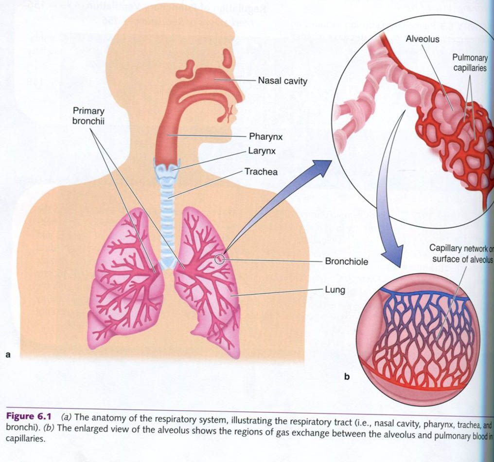
The lungs are not directly attached to the ribs. Rather, they are suspended by the pleural saes. The pleural sacs have a double wall: the parietal pleura, which lines are thoracic wall, and the visceral or pulmonary pleura, which lines the outer aspects of the lung. These pleural walls envelop the lungs and have a thin film of fluid between them that reduces friction during respiratory movements. In addition, these sacs are connected to the lungs and to the inner surface of the thoracic cage, causing the lungs to take the shape and size of the rib or thoracic cage as the chest expands and contracts.
The
anatomy of the lungs, the pleural sacs, and the thoracic cage determines
airflow into and out of the lungs, that is, inspiration and expiration.
Inspiration
Inspiration
is an active process involving the diaphragm and the external intercostales
muscles. Figure a shows the resting positions of the diaphragm and the thoracic
cage, or thorax. With inspiration, the
ribs and sternum are moved by the external intercostal muscles. The ribs swings
up and forward. At the same time, the diaphragm contracts, flattening down
toward the abdomen.
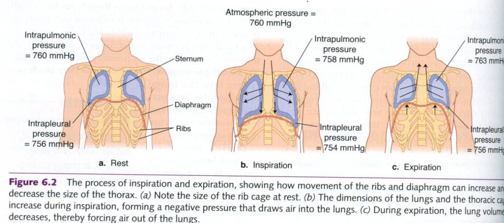
These
actions, illustrated in figure b, expand all three dimensions of the thoracic
cage, increasing the volume inside the lungs. When the lungs are expanded they
have a greater volume, so the air within them has more space to fill. According
to Boyle's gas law, which states that pressure x volume is constant(at a
constant temperature), the pressure within the lungs decreases. As a result,
the pressure in the lungs(intrapulmonary pressure) is less than the air
pressure outside the body. Because the respiratory tract is open to the
outside, air rushes into the lungs to reduce this pressure difference. This is
how air moves into the lungs during inspiration.
During
forced or labored breathing, as during heavy exercise, inspiration is further
assisted by the action of other muscles, such as the scaleni(anterior, middle,
and posterior) and sternocleidomastoid in the neck and the pectoralis in the
chest. These muscles help raise the ribs even more than during regular
breathing.
Expiration
At
rest, expiration is usually a passive process involving relaxation of the inspiratory
muscles and elastic recoil of the lung tissue. As the diaphragm relaxes, it
returns to its normal upward, arched position. As the external intercostal
muscles relax, the ribs and sternum move back into their resting positions(c).
While this happens, the elastic nature of the lung tissue causes it to recoil
to its resting size. This increases the pressure in the lungs and causes a
proportional decrease in volume in the thorax, and therefore air is forced out
of the lungs.
During
forced breathing, expiration becomes a more active process. The internal
intercostales muscle actively pulls down the rib. This action can be assisted
by latissimus dorsi and quadratus lumborum. Contracting the abdominal muscles
increases the intraabdominal pressure, forcing the abdominal viscera upward
against the diaphragm and accelerating its return to the domed position. These muscles also pull the rib
cage down and inward.
The
changes in intra-abdominal and intrathoracic pressure that accompany forced
breathing also help return venous blood back to the heart; this is similar to
the action of the muscle pump in the legs in assisting the return of venous
volume. As intra-abdominal and intrathoracic pressure increases, it is
transmitted to the great veins - the pulmonary veins and superior and inferior
venae cavae - that transport blood back to the heart. When the pressure
decreases, the veins return to their original size and fill with blood. The
changing pressures within the abdomen and thorax squeeze the blood in veins,
assisting in return through a milking action. This phenomenon is known as the
respiratory pump and is essential in maintaining adequate venous return.


0 коментара:
Постави коментар