Extensor digitorum brevis
The lumbricals
The interossei
Extensor digitorum brevis
Extensor digitorum brevis is situated on the
dorsum of the foot beyond the
inferior part of the extensor retinaculum, lateral to and partly covered by the
tendons of peroneus tertius and extensor digitorum longus. It is a thin
muscle arising from the anterior roughened part of the upper surface of the calcaneus
and the deep fascia covering the muscle, including the stem of the inferior
extensor retinaculum. From the small belly, short tendons pass forwards and
medially, the most medial of which crosses the dorsalis pedis artery to insert
separately on to the dorsal aspect of
the base of the proximal phalanx of
the great toe. The remaining three
tendons join the lateral side of the dorsal hood of the second, third and fourth toes. The most medial part of the muscle
may develop a separate belly, sometimes reffered to as extensor hallucis
brevis.
Nerve
supply
This muscle is supplied by the deep peroneal nerve, root value L5, S1.
The skin covering the muscle is supplied by roots L5, S1.
Action
The medial part of the muscle aids extensor hallucis longus in extending
the great toe at the metatarsophalangeal joint, while the other three tendons
aid extensor digitorum longus. As
with the long extensor tendons, extensor digitorum brevis helps the lumbricals
to extend the interphalangeal joints; however, it is unable to do this
independently.
Functional
activity
Extensor digitorum brevis will help extensor digitorum longus and extensor hallucis longus to raise the
toes clear to the ground in running and walking.
Palpation
Place the fingers on the tendon of extensor digitorum longus as it splits
into its four parts. When the toes are extended, extensor digitorum brevis can
be felt just lateral and deep to the tendon. The tendons are difficult to trace
distally as they become inseparable from those of extensor digitorum longus.
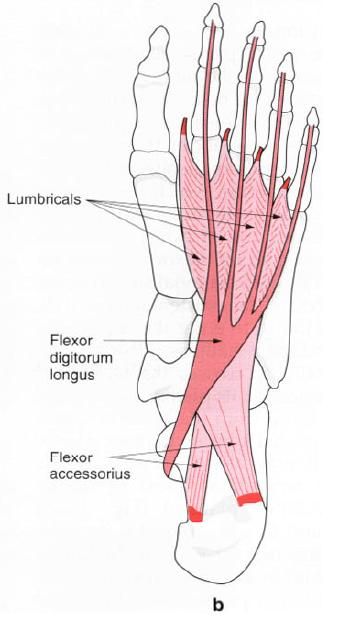
The lumbricals
These are four small muscles associated with
the tendons of flexor digitorum longus; they pass from the flexor to the
extensor compartment of the foot. The
most medial of these muscles arises from the medial side of the tendon
to the second toe, adjacent to the
attachment of flexor accessories (quadratus plantae) to the main longus tendon.
The remaining lumbricals arise by two heads from adjacent sides of two tendons,
that is the second from the tendons to the second and third toes, the third
from the tendons to the third and fourth toes, and the fourth from the tendons
to the fourth and fifth toes. Each muscle then passes forwards below the deep
transverse metatarsal ligament on the medial side of the toe, winding obliquely
upwards to attach to the medial side of the extensor
hood and base of the proximal phalanx.
Nerve
supply
The first and most medial lumbrical is supplied
by the medial plantar nerve, root
value S1, 2, while the lateral three are supplied by the lateral plantar nerve, root value S2, 3; both being terminal
branches of the tibial nerve. The skin on the dorsum of the foot at the point of attachment is supplied by roots L5, S1.
The skin of the plantar aspect of the foot overlying the muscles is supplied by the medial and lateral plantar
nerves, which have the same root values as the supply to the muscles. It should
be noted, however, that only the most lateral of the lumbricals has skin over
its plantar aspect.
Action
There has been much discussion about the role
of these small, almost insignificant muscles. They have a long muscle belly
compared with their tendon and they link the flexors of the toes with the
extensors. By their attachment to the proximal phalanx, contraction of the
lumbricals produces flexion of the toes at the metatarsophalangeal joint.
However, because they also insert into the extensor hood, the lumbricals extend
the interphalangeal joints. Indeed, this latter action is primarily due to the
lumbricals and not the long and short extensor tendons.
Functional
activity
The action of the lumbricals prevents clawing
of the toes in the propulsive phase of gait. Paralysis of these muscles results
in the extensor muscles pulling the toes into hyperextension at the
metatarsophalangeal joint. Even at rest the toes become clawed.
The nerves which supply these muscles appear to
have many more fibres than would be necessary for such a small muscle and a
great number of these are sensory. This leads one to believe that they may have
a very important function in providing information related to the tension
developed between the long flexor and extensor muscles. This sort of
information is of great importance in locomotion, especially as the point of
attachment of the lumbricals is a long way from the muscle bellies of the
extensors and flexors.
Palpation
It is not possible to palpate these muscles as
they lie deep in the sole of the foot
covered by many of the small muscles of the sole and the long flexor tendons.
Dorsal interossei
There are four dorsal interossei, being small
bipennate muscles situated between the metatarsals. Each arises from the proximal half of the sides of adjacent metatarsals, forming a central tendon which passes
forwards, deep to the deep transverse metatarsal ligament. It passes between
the metatarsal heads to attach to the side of the proximal phalanx and capsule of the metatarsophalangeal joint. The
tendons do not attach to the extensor hood.
The first, or most medial, arises from the
adjacent sides of the first and second metatarsals and attaches to the medial
side of the base of the proximal phalanx of the second toe. The second arises
from the adjacent sides of the second and third metatarsals and it also
attaches to the proximal phalanx of the second toe but to the lateral side. The
third and fourth dorsal interossei
attach to the lateral side of the proximal phalanx of the third and fourth toes
respectively.
Nerve
supply
All four dorsal interossei are supplied by the lateral plantar nerve, root value S2, 3,
those in the fourth interosseus space from the superficial branch, and the rest
by the deep branch. The skin covering this area on the dorsum of the foot is supplied by root L5 medially
and S1 laterally.
Action
The dorsal interossei abduct the toes at the
metatarsophalangeal joint, however this action, as such, is of little
importance in the foot. Acting with
the plantar interossei, they will produce flexion of the metatarsophalangeal
joint.
Functional
activity
The dorsal interossei are powerful little
muscles and their activity in combination with the plantar interossei controls
the direction of the toes during violent activity, thus enabling the long and
short flexors to perform their appropriate actions.
These muscles, because of their relationship to
the metatarsophalangeal joint, can flex these joints and so raise the heads of
the second, third and fourth metatarsals, thus helping to maintain the anterior
metatarsal arch. They also help, to a limited extent, with the maintenance of
the medial and lateral longitudinal arches of the foot.
Palpation
Place the finger tips between the proximal
parts of the metatarsals on the dorsum of the foot; when the toes are abducted; the muscles can be felt to contract.

Plantar interossei
The plantar interossei are smaller than their
dorsal counterparts, fusiform in shape and found in the lateral three
interosseus spaces. Each arises from the plantar
and medial aspect of the base and proximal end of the shaft
of the metatarsal. The tendon formed
passes forwards and deep to the deep transverse metatarsal ligament to insert
into the medial side of the base of the proximal phalanx of the same
toe.
Nerve
supply
All the interossei are supplied by the lateral plantar nerve, root value S2, 3,
with that in the fourth interosseus space being supplied by the superficial
branch of the nerve. The skin covering the area is supplied on the lateral side
by root S1 and medially by root L5.
Action
The plantar interossei adduct the third, fourth
and fifth toes towards the second. In conjunction with the dorsal interossei
they flex the metatarsophalangeal joints of the lateral three toes.
Functional
activity
With the help of the dorsal interossei and
abductor digiti minimi, the plantar interossei help to control the position of
the third, fourth and fifth toes during the push-off phase of walking and
running. They also help to prevent splaying of the toes when weight is suddenly
applied to the forefoot.
Palpation
These muscles are too deep to be palpated.