


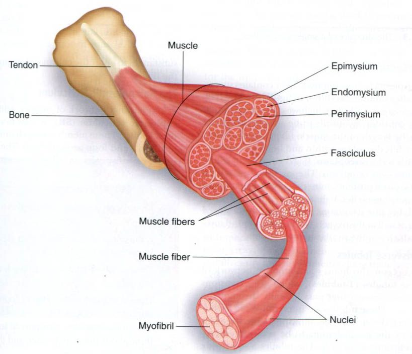
If a person were to dissect a muscle, he or she
would first cut through the outer connective tissue covering. This is the epimysium. It surrounds the entire
muscle, holding it together. Once one had cut through the epimysium, one would
see small bundles of fibers wrapped in a connective tissue sheath. These
bundles are called fasciculi. The connective tissue sheath surrounding each fasciculus is the perymysium.
Finally, by cutting through the perimysium and
using a microscope, one would see the muscle
fibers, which are the individual muscle cells. A sheath of connective
tissue, called the endomysium, also
covers each muscle fiber. It is generally thought that muscle fibers extend
from one end of the muscle to the other; but under the microscope, muscle
bellies often divide into compartments or more transverse fibrous
bands(inscriptions). Because of this compartmentalization, the longest human
muscle fibers are about 12cm, which corresponds to about 500,000 sarcomeres,
the basic functional unit of the myofibril. The number of fibers in different
muscles ranges from several hundred(e.g. tensor tympani, attached to the
eardrum) to more than a million(e.g. medial gastrocnemius muscle).
Muscle Fiber
Muscle fibers range in diameter from 10 to 120 µm, so they are nearly invisible to the naked
eye. The following sections describe the structure of the individual muscle
fiber.
Plasmalemma
If one looked closely at an
individual muscle fiber, one would see that it is surrounded by a plasma membrane,
called the plasmalemma. The
plasmalemma is part of a larger unit reffered to as the sarcolemma. The sarcolemma is composed of the plasmalemma and the
basement membrane. Some textbooks use the term sarcolemma to describe just the
plasmalemma. At the end of each muscle fiber, its plasmalemma fuses with the
tendon, which inserts into the bone. Tendons are made of fibrous cords of
connective tissue that transmit the force generated by muscle fibers to the
bones, thereby creating motion. So typically, individual muscle fibers are
ultimately attached to bone via the tendon.
The plasmalemma has several
unique features that are important to muscle fiber function. It appears as a
series of shallow folds along the surface of the fiber when the fiber is
contracted or in a resting state, but these folds disappear when the fiber is
stretched. This folding allows stretching of the muscle fiber without
disrupting the plasmalemma. The plasmalemma also has junctional folds in the
innervation zone at the motor end plate, which assists in the transmission of
the action potential from the motor neuron to the muscle fiber as discussed
later in this chapter. Finally, the plasmalemma helps to maintain acid-base
balance and transports metabolites from the capillary blood into the muscle
fiber.
Satellite cells are located between the plasmalemma and the basement membrane. These
cells are involved in the growth and development of skeletal muscle and in
muscle’s adaptation to injury, immobilization, and training.
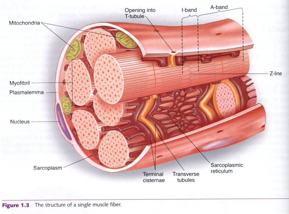
Sarcoplasm
Inside the plasmalemma, a muscle
fiber contains successively smaller subunits, as shown in the figure. The
largest of these are myofibrils, which we discuss separately. For now, consider
myofibrils to be rodlike structures running the length of the muscle fibers. A
gelatin-like substance fills the spaces within and between the myofibrils. This
is the sarcoplasm. It is the fluid
part of the muscle fiber – its cytoplasm. The sarcoplasm contains mainly
dissolved proteins, minerals, glycogen, fats and the necessary organelles. It
differs from the cytoplasm of most cells because it contains a large quantity of
stored glycogen as well as the oxygen-binding compound myoglobin, which is
quite similar to hemoglobin.
The Transverse Tubules
The sarcoplasm also houses an
extensive network of transverse
tubules(T-tubules), which are extensions of the plasmalemma that pass
laterally through the muscle fiber. These tubules are interconnected as they
pass among the myofibrils, allowing nerve impulses received by the plasmalemma
to be transmitted rapidly to individual myofibrils. The tubules also provide
pathways from outside the fiber to its interior, enabling substances to enter
the cell and waste products to leave the fibers.
The Sarcoplasmic Reticulum
A longitudinal network of
tubules, known as the sarcoplasmic
reticulum(SR), is also found within the muscle fiber. These membranous
channels parallel the myofibrils and loop around them. The SR serves as a storage
site for calcium, which is essential for muscle contraction. Figure shown
depicts the T-tubules and the SR.


0 коментара:
Постави коментар