


Striations and the Sarcomere

Under a light microscope, skeletal muscle
fibers have a distinctive striped appearance. Because of these markings,
skeletal muscle is also called striated muscle. This striation also is seen in
cardiac muscle, so it too can be considered striated muscle.
As you can see in the picture, you will see the
color difference between A-bands, and I-bands. Each dark A-band has a
lighter region in its center, the H-zone, which is visible only when myofibril
is relaxed. There is a dark line in the middle of H-zone called the M-line.
I-bands are interrupted by a dark stripe referred to as the Z-disk, also known
as the Z-line.
A sarcomere
is the basic functional unit of a myofibril and the basic contractile unit of
muscle. Each myofibril is composed of numerous sarcomeres joined end to end at
the Z-disks. Each sarcomere includes what is found between each pair of
Z-disks, in this sequence:
- An
I-band(light zone)
- An
A-band(dark zone)
- An
H-zone(in the middle of the A-band)
- An
M-line in the middle of the H-zone
- The
rest of the A-band
- A
second I-band
Looking at individual myofibrils through an
electron microscope, one can differentiate two types of small protein filaments
that are responsible for muscle contraction. The thinner filaments are composed
primarily of actin, and the thicker
filaments are primarily myosin. The
striations seen in muscle fibers result from alignment of these filaments. The light I-band indicates the region of
the sarcomere where there are only thick filaments. The dark A-band represents the regions
that contain both thick and thin filaments. The H-zone is the central portion of the A-band and contains
only thick filaments. The absence of thin filaments causes the H-zone to
appear lighter than the adjacent A-band. In the center of the H-zone is the M-line, which is composed of proteins
that serve as the attachment site for the thick filaments and assist in
stabilizing the structure of the sarcomere. Along with two additional
proteins, titin and nebulin, they provide points of
attachment and stability for the thin filaments.
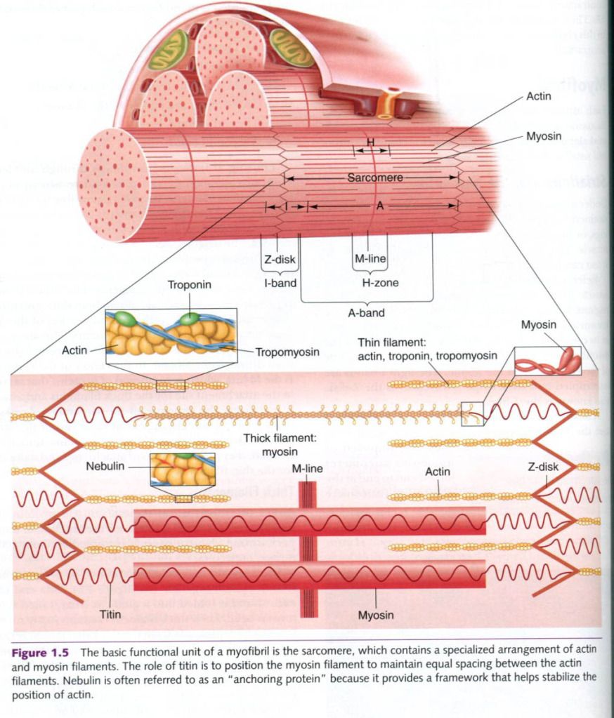
Thick
filaments
About two-thirds of all skeletal muscle protein
is myosin, the principal protein of the thick filament. Each myosin filament
typically is formed by about 200 myosin molecules.
Each myosin molecule is composed of two protein
strands twisted together. One end of each strand is folded into a globular
head, called the myosin head. Each thick filament contains many such heads,
which protrude from the thick filament to form cross-bridges that interact
during muscle contraction with specialized active sites on the thin filaments.
There is an array of fine filaments, composed of titin, that stabilizes the myosin filaments along their
longitudinal axis. Titin filaments extend from the Z-disk to the M-line.
Thin
filaments
Each thin filament, although often reffered to
simply as an actin filament, is actually composed of three different protein
molecules – actin, tropomyosin, and
troponin. Each thin filament has one end inserted into a Z-disk with the
opposite and extending toward the center of the sarcomere, lying in the space
between the thick filaments. Nebulin,
an anchoring protein for actin, coextends with actin and appears to play a
regulatory role in mediating actin and myosin interactions(see in picture). Each
thin filament contains active sites to which myosin heads can bind.
Actin forms the backbone of the filament.
Individual actin molecules are globular proteins(G-actin) and join together to
form strands of actin molecules. Two strands then twist into a helical pattern,
much like two strands of pearls twisted together.
Tropomyosin is a tube-shaped protein that
twists around the actin strands. Troponin is a more complex protein that is
attached at regular intervals to both the actin strands and the tropomyosin. Tropomyosin
and troponin work together in an intricate manner along with calcium ions to
maintain relaxation or initiate contraction of the myofibril.
“Physiology of sport and exercise”, fourth edition; Jack H. Wilmore, David L. Costill, W. Larry Kenney
“Physiology of sport and exercise”, fourth edition; Jack H. Wilmore, David L. Costill, W. Larry Kenney


0 коментара:
Постави коментар