


The ulna lies medial to the radius and is the
longer of the two bones. It has a shaft and two ends, of which the superior is
the larger presenting as a hook-like projection for articulation with the
trochlea of the humerus. The smaller rounded distal end is the head of the
ulna. It does not articulate directly with the carpus. The ulna articulates
laterally at each end with the radius.
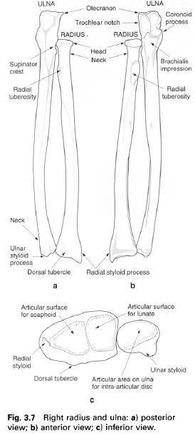
The upper end of the ulna is large with two
projecting processes, enclosing a concavity. The olecranon process is the larger of the two processes and forms the
proximal part of the bone. It is beak-shaped and points forwards, being
continuous inferiorly with the shaft. Posteriorly, it is smooth and
subcutaneous while anteriorly it is concave, forming the upper part of the
articular surface of the trochlear notch.
The borders of the olecranon are thickened and rough.
The coronoid
process projects from the front of the shaft and has an upper articular
surface which completes the trochlear notch. These two surfaces are often
separated by a roughened non-articular area running horizontally across the
notch. The trochlear notch is divided by a vertical ridge into a larger medial
part and a smaller lateral part, the latter being continuous over its outer
edge with the articular surface of the radial notch on the lateral side of the
coronoid process. There is a small tubercle where the medial and anterior edges
of the articular surface of the coronoid process meet. This gives attachment to
the anterior part of the ulnar collateral ligament. The irregular, anterior
surface of the coronoid ends inferiorly as the rough tuberosity of the ulna.
Both this surface and the tuberosity give attachment to brachialis. At the
upper medial part of the coronoid is the small sublime tubercle from which the
pronator ridge runs downwards and laterally.
On the lateral side of the coronoid process,
the concave radial notch receives the head of the radius. Below this and
extending onto the shaft is the triangular supinator fossa. It is bound
posteriorly by the distinct supinator
crest. The medial border of this area forms a prominent ridge which has a
small tubercle at its upper end.
The prominent interosseus border, to which the
interosseous membrane attaches, runs down from the apex of the supinator fossa.
The anterior border runs down from the medial margin of the coronoid process
but is indistinct. The sinuous, subcutaneous posterior border, prominent in its
upper part, is continuous with the subcutaneous region of the olecranon and
upper part of the shaft. Between these borders are three surfaces, the anterior
and medial surfaces being continuous at the rounded anterior border. The lower
quarter of the anterior surface is marked by an oblique ridge running downwards
and medially. On the posterior surface, an oblique ridge runs downwards and
backwards from the radial notch to the posterior border. The remaining
posterior surface has faint ridges laterally and is smooth medially.
The lower end of the ulna shows a narrowed neck
which expands into a small, rounded head. From the posteromedial part of the
head of the ulna, the conical styloid
process projects downwards. The head has a smooth articular surface for the
radius on its anterior and lateral aspects. The distal surface of the head is
smooth and almost flat, and articulates with an articular disc which intervenes
between it and the triquetral.
Palpation
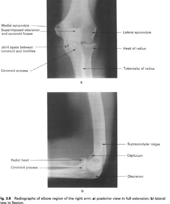
At the upper and posterior aspect of the elbow
the outline of the olecranon can be identified; it forms the “point” of the
elbow seen in flexion. Running downwards from this point the posterior border
can be palpated throughout its length. At the lower end, the neck, head and
styloid process can be palpated, with the styloid process being the most
posterior. When the forearm is fully pronated the rounded head of the ulna
stands out from the back of the wrist.
Ossification
A primary ossification centre appears in the
shaft during the eighth week in utero. The body, coronoid process and major
part of the olecranon ossify from this primary centre. A secondary centre
appears in the head during the fifth year and fuses with the shaft between 20
and 22 years. The secondary centre for the remainder of the olecranon appears
at about 11 years, with fusion occurring between 16 and 19 years. There may be
several secondary centres for the olecranon.


0 коментара:
Постави коментар