


Rhomboid minor
Rhomboid major
Trapezius
Rhomboid minor
Rhomboid minor is a small quadrilateral muscle
whose fibres run obliquely downwards and laterally from the spinous processes of C7 and T1 and the supraspinous ligament between them and
the lower part of the ligamentum nuchae, to attach the medial border of the smooth triangular
area at the base of the spine of the
scapula.
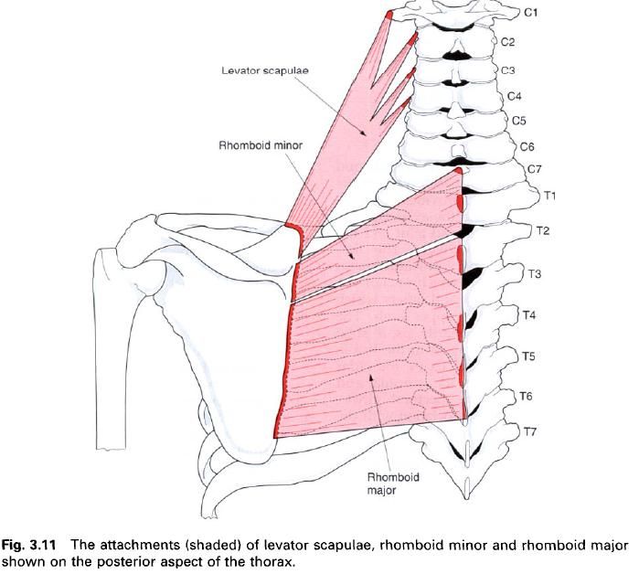
Rhomboid major
Rhomboid major, although larger than rhomboid
minor, may be continuous with it. It arises by tendinous slips from the spinous processes of T2 to T5 inclusive
and the intervening supraspinous ligament.
The muscle fibres run obliquely downwards and laterally to attach to a medial border of the scapula between the base of the spine
and the inferior angle.
Both rhomboids lie superficial to the long back
muscles, being themselves covered by trapezius, except for the lower border of
rhomboid major which forms the floor of the “triangle of auscultation”.
Nerve
supply
Both rhomboid muscles are supplied by the dorsal scapular nerve, root value C5.
Action
The rhomboids act principally to retract the scapula but are also active, however, in
medial rotation of the pectoral girdle. In addition they also act as important
stabilizers of the scapula when other
muscle groups are active.
Palpation
With the subject’s hand placed in the small of
the back(to relax trapezius), the rhomboids can be palpated through trapezius
when the hand is moved backwards. Contraction of the rhomboids can be felt(and
occasionally be seen) between the medial border of the scapula and the vertebral column.
Trapezius
Trapezius is a large, flat triangular sheet of
muscle extending from the skull and spine medially to the pectoral girdle
laterally. It is the most superficial muscle in the upper back and with its
fellow of the opposite side it forms a trapezium, hence its name.

The medial attachment of trapezius runs from
the medial third of the superior nuchae
line and external occipital protuberance
of the occipital bone, the ligamentum
nuchae, the spinosus processes of
C7 to T12 inclusive and the intervening supraspinous
ligament. The majority of this attachment is by direct muscular slips,
however a triangular aponeurosis exists in trapezius between C6 and T3 which
corresponds to a hollow seen in the living subject.
From this extensive central attachment the
upper fibres of trapezius run downwards and laterally, the middle fibres almost
horizontally, and the lower fibres upwards and laterally to form a continuous
line of attachment to the clavicle
and scapula. The upper fibres descend
to the posterior border of the lateral
third of the clavicle, while the
middle fibres pass to the medial border
of the acromion and upper border of the crest of the spine of the
scapula, being separated from the smooth area on the medial part of the
spine by a small bursa. The lower-most fibres converge to a tendon which
attaches to the tubercle on the inferior edge at the medial end of the spine of the scapula.
The upper free edge of trapezius forms the
posterior border of the posterior triangle of the neck, while the lower free
border forms the medial boundary of the triangle of auscultation. This latter
triangle is an area of the chest wall free of bony obstruction by the scapula
and thinly covered by muscle. Its other boundaries are the upper border of
latissimus dorsi below and the medial border of the scapula laterally.
Nerve
supply
Trapezius receives its motor supply via the
spinal part of the accessory nerve(XI)
which enters it from the posterior triangle. It also receives sensory fibres
from the ventral rami of C3,4 via the
cervical plexus. The skin over trapezius is supplied by the dorsal rami of C3 – T12.
Action
Trapezius has an important function in
stabilizing the scapula as a base for
movements of the upper limb. The middle horizontal fibres pull the scapula backwards towards the midline,
that is retraction, and may be aided
by the upper and lower fibres contracting together to produce a “resolved”
force towards the midline. The upper fibres of trapezius elevate the pectoral
girdle and maintain the level of the shoulders against the effect of gravity,
or when a weight is being carried in the hand. When both left and right muscles
contract they can extend the neck, but when acting singularly the upper fibres
produce lateral(side) flexion of the neck. The lower fibres pull down the
medial part of the scapula and thus lower the shoulder, especially against
resistance, for example when using the arms to get out of a chair. The upper
and lower fibres working together produce lateral rotation of the scapula about a
point towards the base of the spine. Trapezius is thus important in the overall
function of the upper limb as its action increases the possible range of
movement.
Paralysis of trapezius, particularly its upper
part, results in the scapula moving forwards around the chest wall with the
inferior angle moving medially. The usually smooth curve of its upper border
between the occiput and the acromion process may become markedly angulated.
Palpation
To demonstrate and palpate all three parts of
trapezius, the subject should abduct both arms to 90°, flex the elbows to 90°
and then rotate them laterally so that the fingers are pointing upwards.
In this position the three sets of fibres can
be readily palpated; in a lean subject the contraction of the various parts of
the muscle can be seen. For the lower fibres of trapezius the contraction can
be further enhanced by asking the subject to clasp his or her hands together
above the head and pull hard.
Soft tissue techniques are often applied to the
upper muscular fibres of trapezius in the presence of muscle spasm secondary to
neck pain, with the aim of inducing relaxation. Deep transverse frictions can also
be applied to the tendinous attachment of trapezius on the superior nuchal line
when this is the site of a lesion causing pain in the neck or occipital region.


1 коментара:
Love this right here!
Trapezius Stretches
Постави коментар