


The metacarpus
The metacarpus consists of five bones, the
metacarpals, one corresponding to each digit and numbered in sequence from the
lateral side. Each is a long bone with a proximal quadrilateral base, a shaft
and a distal rounded head. The
variations in shape of the ases provide a means of distinguishing them. The
base of the first metacarpal has a saddle-shaped articular surface which fits a
corresponding surface on the trapezium. The base of the second metacarpal
articulates with the trapezium, trapezoid and capitate. The base of the third
has a single articulation with the capitate. The bases of the fourth and fifth
metacarpals articulate with the hamate. The bases of the second to fifth also
articulate with the adjacent metacarpal bones, having articular facets in
appropriate positions.
The heads of the metacarpals are smooth and
rounded, extending further onto the palmar surface. The palmar articular margin
is notched in the midline. The head of the first metacarpal is wider than the
others having two sesamoid bones,
usually found in the short tendons crossing the joint and which articulate with
the palmar part of the joint surface occasionally grooving it. The heads fit
into a concavity on the base of the proximal phalanx at the metacarpophalangeal
joints. The shaft of the metacarpals is slightly curved with a longitudinal
palmar concavity. That of the first metacarpal is nearly as wide as the base
and has a rounded dorsal surface. The palmar surface is divided by a blunt
ridge into a larger lateral part and a smaller medial part.
Palpation
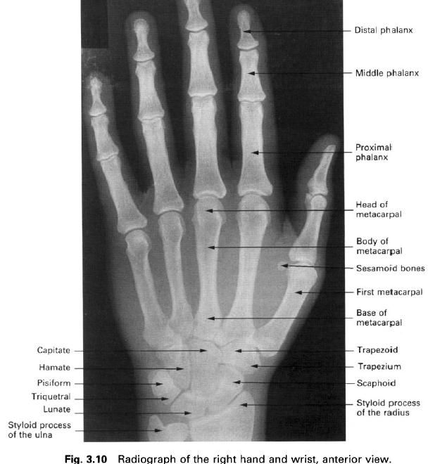
If the fingers are flexed to form a fist, the
heads of the metacarpals can easily be palpated as the knuckles. Running
proximally on the dorsal surface of the hand the shafts can also be
distinguished. At the proximal end of the shaft the gap between the base of the
metacarpal and the carpus can be palpated as the line of the carpometacarpal
joint.
Ossification
Primary ossification centres appear in the
shaft in the ninth week in utero, so
that the bones are well ossified at birth. Secondary centres appear in the
heads of the second to fifth metacarpals between 2 and 3 years. The secondary
centre for the base of the first metacarpal appears slightly later. Fusion of
the epiphysis with the shaft occurs between 17 and 19 years for all
metacarpals. Occasionally, a secondary centre may appear in the head of the
first metacarpal.
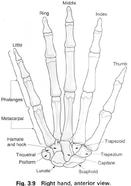
The phalanges
There are 14 phalanges in each hand, three for
each finger and two for the thumb. As they are long bones, each phalanx has a
shaft, a large proximal end and a smaller distal end, the head. The phalanges
of the thumb are shorter and broader than those of the fingers.
The proximal phalanx has a concave oval facet
on its base for articulation with the head of the metacarpal. The rounded head,
which extends further onto the palmar surface, has a wide, pulley – shaped
articular surface for the base of the next phalanx. The shaft is curved along
its length being convex dorsally. It is convex from side to side on its dorsal
surface, and is flat on the palmar surface. The middle and distal phalanges are
similar to the proximal phalanx. However, the base of the distal phalanx is
large, and the head is expanded to support the pulp pad of the digits.
By convention, the digits are described by name
rather than by number, and are from lateral to medial, the thumb, index,
middle, ring and little fingers.
Palpation
By flexing the fingers into a fist, the heads
of the proximal and middle phalanges can be palpated. The shafts of the
phalanges are also easily followed throughout their length, especially on their
dorsal surface.
Ossification
Primary ossification centres appear in the
shafts of the phalanges between the eighth and twelfth week in utero, with the
distal phalanges ossifying first. Secondary centres appear in the bases of the
phalanges during the second and third year, fusing with the shaft between 17
and 19 years. Occasionally, a secondary centre may appear in the head as well
as in the base.


0 коментара:
Постави коментар