


Glands
Sebaceous glands
These are associated with all hairs and hair
follicles, there being between one and
four associated with each hair. They may also exist where there is no hair,
such as the corner of the mouth and adjacent mucosa, the lips, the areola and
the nipple, opening directly onto the skin surface. However, they are absent
from the skin of the palm and sole and the dorsum of the distal segments of the
digits. The glands vary in size between 0.2 and 2.0mm in diameter. The cells of
the glands are continuously destroyed in the production of the oily secretions,
known as sebum. This mode of secretion production is known as holocrine secretion.
Inflammation and accumulation of secretion
within the sebaceous glands give rise to acne. If plugging of the outlet is
permanent, a sebaceous cyst may be formed in the duct and follicles. These may
become so enlarged that they require surgical removal. Sebaceous glands do not
appear to be under nervous control.
Sweat glands
These have a wide distribution throughout the
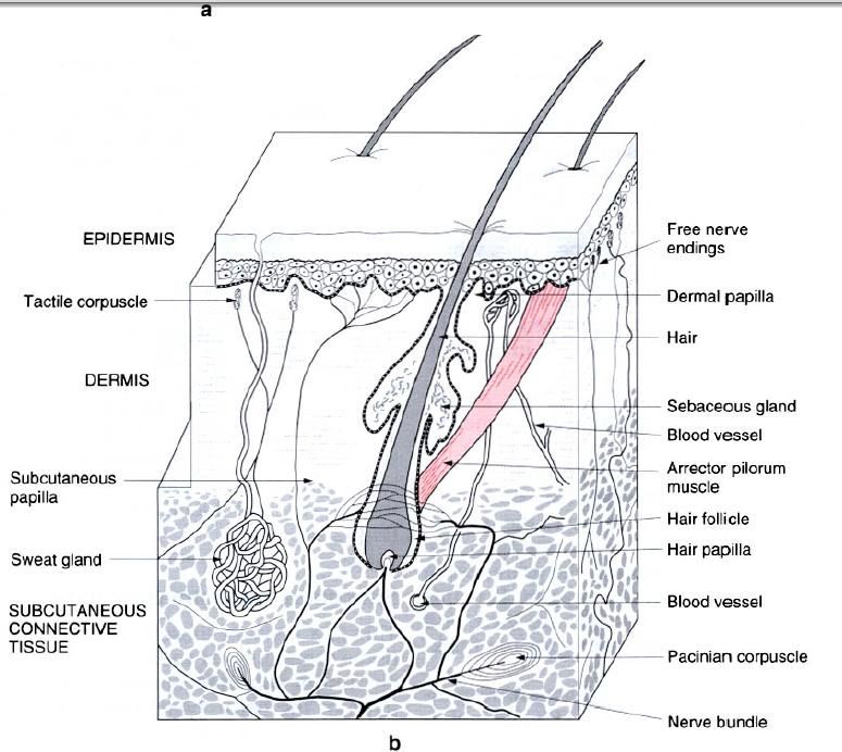
body(figure below), being more numerous on its exposed parts, especially on the
palms, soles and flexor surfaces of the digits. Here the ducts open onto the
summits of the epidermal ridges. Each gland has a long tube extending into the
subcutaneous tissue, where it becomes coiled forming the secretory body of the
gland(figure b). The glands produce sweat, which is a clear fluid without any
cellular elements, for secretion(eccrine
secretion). The production of sweat is important in temperature regulation,
as its evaporation from the skin surface promotes heat loss. These eccrine
sweat glands are quently, any disturbance in the sympathetic system will result
in a dry warm skin(anhydrosis) either locally or extensively.
In the axilla, groin and around the anus there
are large modified sweat glands, being between 3 and 5mm in diameter and lying
deeply in the subcutaneous layer. Their ducts may be associated directly onto
the skin surface. The secretions of the glands include some disintegration
products of the gland cells(apocrine
secretions). The odour associated with these glands is not from the
secretion itself, but is due to bacterial invasion and contamination from the
skin. Pigment granules associated with axillary glands produce a slight
coloration of the secretion. The apocrine glands vary with sexual development,
enlarging at puberty. In females they show cyclical changes associated with the
menstrual cycle.
The glands which open at the margins of the
eyelid(ciliary glands) are modified,
uncoiled sweat glands, as are the glands of the external auditory meatus(ceruminous glands). The cells of these
latter glands contain a yellowish pigment which colours the wax secretion(cerumin).
Mammary gland(breast)
The mammary glands are modified sweat glands,
being accessory to the reproductive function in females, secreting
milk(lactation) for the nourishment of the infant. In children prior to puberty
and the adult male, the glands are rudimentary and functionless.
Blood
supply and lymphatic drainage of the skin
The arterial supply of the skin is derived from
vessels in the subcutaneous connective tissue layer, which form a network at
the boundary between the dermis and subcutaneous tissue(figure b). Branches
from the network supply the fat,
sweat glands and deep parts of the hair follicles. Branches within the dermis
form a subpapillary plexus. The epidermis is avascular. Abundant arteriovenous
anastomoses occur within the skin. Lymphatics of the skin begin in the dermal
papillae as networks or blind outgrowths which form a dense mesh of lymphatic
capillaries in the papillary layer. Larger lymphatic vessels pass deeply to the
boundary between dermis and subcutaneous tissue to accompany the arteries as
they pass centrally.
Nerves
of the skin
The nerves of the skin are of two types,
afferent somatic fibres mediating pain, touch, pressure, heat and cold(general
sensations), and efferent autonomic(sympathetic) fibres supplying blood
vessels, arrector pilorum and sweat glands. The sensory(afferent) endings have
several forms. Free nerve endings extend between cells of the basal layer of
the epidermis, terminating around the adjacent to hair follicles. They are
repetitive to general tactile sensation as well as painful stimuli. Enclosed
tactile corpuscules lie in the dermal papillae, being sensitive to touch.
Pacinian corpuscules(figure b) exist in the subcutaneous tissue, being
particularly plentiful along the sides of the digits, and act as pressure
receptors. Specific endings for heat and cold have been described, although general
agreement as to their identity has not been reached.


0 коментара:
Постави коментар