


The radius lies lateral to the ulna and is the
shorter of the two bones. It articulates above with the capitulum of the
humerus, distally with the scaphoid and lunate bones of the proximal row of the
carpus, and at each end with the ulna. It has a shaft and two ends, the
inferior end being the larger.
The head
is a thick disc with a concave superior surface for articulation with the
capitulum. The outer, articular surface of the head is flattened, articulating
inside a fibro-osseous ring formed by the radial notch of the ulna and the
annular ligament. Below the head is the constricted neck, which slopes medially as it approaches the shaft. Where the
shaft joins the neck it is round, but becomes triangular lower down. Together
with the neck, the shaft has a slight medial convexity on its upper quarter,
with a lateral convexity in its remaining lower part. The radial tuberosity lies anteromedially on the upper part of the
shaft at the maximum convexity of the medial curve. The majority of the shaft
presents three borders and three surfaces.
The interosseous border, to which the
interosseous membrane attaches, is sharp and faces medially. It extends from
just below the radial tuberosity to the medial side of the lower end of the
radius, splitting into two ridges which become continuous with the anterior and
posterior margins of the ulnar notch. The anterior and posterior borders pass
obliquely downwards and latteraly from either side of the radial tuberosity to
the roughened area for pronator teres lower down, while the posterior border
becomes more rounded. These borders enclose the lateral, anterior and flatter
posterior surfaces.
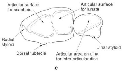
The inferior end of the radius is expanded
having five distinct surfaces. The lateral surface, which extends down to the
styloid process, has a shallow groove anteriorly for the tendons of abductor
pollicis longus and extensor pollicis brevis. The medial surface forms the
concave ulnar notch for articulation with the head of the ulna having a
roughened triangular area supeiorly. The posterior surface is convex and
grooved by tendons. The prominent ridge in the middle of this surface is the
dorsal(Lister’s) tubercle. The lateral half of this surface continues down onto
the styloid process. The anterior surface is smooth and curves forward to a
distinct anterior margin. The distal articular surface is concave and extends
onto the styloid process. It is divided by a ridge into two areas, a lateral
triangular area for articulation with the scaphoid and a medial quadrilateral
area for the lunate.
Palpation
The head of the radius can be palpated in a
“dimple” on the posterolateral aspect of the elbow, particularly when the elbow
joint is extended as it overhangs the capitulum; it can be felt to rotate
during pronation and supination. The shaft of the radius can be palpated on the
lateral side in the lower half of the forearm. Distally, on the posterior
aspect, the dorsal tubercle can be identified above the wrist, as can the
styloid process laterally inside the “anatomical snuff-box” between the
extensor tendons of the thumb.
Ossification
A primary ossification centre appears in the
shaft during the eighth week in utero, so that at birth only the head, inferior
end and radial tuberosity are cartilaginous. The first secondary centre appears
in the inferior end during the first year of life, fusing with the shaft
between the ages of 20 and 22 years. The secondary centre for the head appears
at about 6 years and fuses with the shaft between 15 and 17 years. A secondary
centre usually appears in the radial tuberosity between 14 and 15 years, but
soon fuses with the shaft.


0 коментара:
Постави коментар