


Development of ear
The ear consists of three parts which function
together but have different origins. The membranous part of the internal ear
originates from the otic vesicle(surface ectoderm origin) during the fourth
week. The otic vesicle divides into an anterior part which forms the saccule
and the cochlear duct, and a dorsal part forming the utricle, semicircular
canals and endolymphatic duct. The surrounding bony labyrinth develops from
adjacent mesenchyme. Except for the cochlear duct, from which develops the
organ of Corti(spiral organ), the membranous labyrinth is concerned with
maintaining balance.
The
epithelial lining of the middle ear(tymphanic cavity, mastoid antrum and auditory) tube is derived from the
endoderm of the tubotympanic recess of the first branchial pouch. The auditory
ossicles develop from the dorsal ends of the cartilages of the first(malleus
and incus) and second(stapes) branchial arches.
The
external auditory(acoustic)
meatus develops from the first branchial cleft, and is separated from the
tympanic cavity by the tympanic membrane, which is derived from three sources:
the ectoderm of the first branchial cleft, an intermediate mesodermal layer,
and the endoderm of the first branchial pouch. The external ear(auricle) develops from six mesenchymal swellings
around the margin of the first branchial pouch.
Components of ear
The three parts of the ear(external, middle and internal) are all, except for the auricle,
found within the temporal bone; the auricle is attached to the tympanic
part of the temporal bone. The external ear collects the sounds and conveys
these to the tympanic membrane causing it to vibrate. The tympanic membrane forms the boundary between the external and
middle parts of each ear. Vibration of this membrane is transmitted across the
middle ear by the three auditory
ossicles(incus, malleus and stapes)
to the internal ear. The middle ear communicates with the nasopharynx via the Eustachian(auditory) tube. The internal
ear consists of two functionally distinct parts, that concerned with
hearing(the cochlear part) and that with balance and position(the vestibular
part). The sensory endings of both parts are supplied by the vestibulocochlear
nerve, the eighth cranial nerve.
External ear
The external ear consists of the auricle and the external acoustic(auditory) meatus(see figure) which collect and
convey sound respectively towards the tympanic membrane. The auricle projects
backwards and laterally from the side of the head, being connected to the
fascia by three small, insignificant muscles. It is a single piece of elastic
cartilage, except for the fibrofatty lobule, covered with skin; the named parts
are given in figure a. In adults its shape is extremely variable, increasing
threefold in length from birth to adulthood: it also tends to increase in size
and thickness in old age.
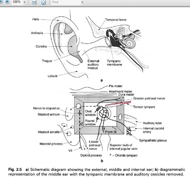
The external acoustic meatus is 25mm long. It
is cartilaginous in its outer third, being continuous with the cartilage of the
auricle, and bony in its medial two thirds, being formed by the tympanic part
of the temporal bone. The meatus curves upwards and backwards as it passes
medially, its inferior wall being 5mm longer than the superior wall because of
the obliquity of the tympanic membrane. The skin lining the meatus is firmly
attached to the underlying bone and in the outer third of the canal contains
numerous ceruminous(wax secreting) cells and hairs. The meatus lies behind the
temporomandibular joint, with the mastoid air cells being immediately
posterior.
Middle ear
This is a narrow, irregular cavity containing
the auditory ossicles immediately medial to the tympanic membrane(figure a).
The middle ear can be conveniently viewed as a six-sided space, with that part
above the tympanic membrane being known as the epitympanic recess. The cavity
communicates with the nasopharynx via the Eustachian tube which opens into the
anterior wall, and with the mastoid air cells via the aditus in the posterior
wall(figure b). The auditory tube enables the pressure on both sides of the tympanic
membrane to be equalized; it is opened during swallowing.
The tympanic membrane is circular and concave
laterally, and consists of three layers: modified skin externally, mucous
membrane internally with an intermediate fibrous layer. The majority of the
membrane is tense, however a small flaccid area exists anterosuperiorly.
Between the internal and external layers runs the chorda tympani branch of the
facial nerve conveying taste sensations from the anterior two thirds of the
tongue.
The auditory ossicles articulate by synovial
joints and transmit the vibrations of the tympanic membrane to the inner ear. The
malleus attaches to the inner surface of the membrane and articulates with the
incus, which in turn articulates with the stapes, the oval base of which lies
in the oval window. Movements of the malleus and stapes are contolled and reflexly
dampened down by contraction of the tensor tympani and stapedius muscles
respectively, both of which are found within the middle ear. Tensor tympani is
innervated by the mandibular division of the trigeminal nerve and stapedius by
the facial nerve.
Internal ear
This is situated within the petrous part of the
temporal bone and consists of a complex series of fluid – filled spaces, known
as the membranous labyrinth, occupying a similarly shaped cavity, the bony
labyrinth. Displacement of the fluid in these spaces stimulates the sensory
endings of the lining epithelium.
The bony labyrinth consists of three parts, the
vestibule(containing the utricle and saccule of the membranous labyrinth), the
semicircular canals(anterior, posterior and lateral) and the cochlea(figure
below). The anterior and posterior semicircular canals are at right angles to
each other and lie 45 degrees to the sagittal plane, with the anterior being
anterior and lateral, and posterior, posterior and lateral. The membranous
semicircular ductus are dilated at one end(the ampulla) in which there is a
thickening(the ampullary crest) where endings of the vestibulocochlear nerve
terminate. The three ducts open into the utricle, which communicates with the
saccule, which in turn communicates with the cochlea. Thickenings in both the
utricle and saccule are known as the maculae and contain terminations of the
vestibulocochlear nerve. The ampullary crests of the semicircular canals convey
information about rotatory and angular movements of the head, while the maculae
convey information regarding linear and tilting movements. Disease of the
semicircular ducts, utricle and saccule gives rise to giddiness of varying
degrees.

The bony cochlea consists of two and
three-quarter turns of a spiral, and resembles a shell lying on its side. It
has a central supporting column of bone(the modiolus) to which is attached a
thin lamina of bone partially dividing the spiral into two parts, the scala
vestibule above and scala tympani below(picture b up). The membranous cochlear duct lines the bony
cochlea and is triangular in cross section. The outer wall of the triangle is
thickened to form the spiral ligament, the lower part is the basilar membrane
while the upper part is the vestibular membrane. The thickened and highly
specialized spiral organ(organ of Corti) lies on the basilar membrane.
Pulsations transmitted to the perilymph within the membranous cochlea by movement
of the stapes in the oval window pass through the scala tympani, and are
transmitted to the fluid in the scala vestibule, being adjusted by compensatory
movements of the round window, thus causing movement of the basilar membrane,
thereby stimulating the hair cells of the spiral organ(picture c); the end
result being auditory perception. Low frequency sounds cause maximum activity
in the basilar membrane; high frequency sounds are limited to the basal portion
of the cochlea.


0 коментара:
Постави коментар