


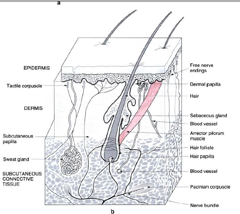
These are nails, hairs, sebaceous, sweat and
mammary glands, and are all derived from the epidermis.
Nails
The nail consists of an approximately
rectangular plate of horny tissue found on the dorsum of the terminal phalanx
of the fingers, thumb and the toes. They are a special modification of the two
most superficial layers of the epidermis, particularly the stratum lucidum. Its
transparency allows the pinkness of the underlying highly vascular nail bed to
show through. The nail is partly surrounded by a fold of skin, the nail wall,
and is firmly adherent to the underlying highly vascular nail bed with some
fibres ending in the periosteum of the distal phalanx. It is this firm
attachment which enables the nails to be used for scratching and as instruments
for prizing open various objects.
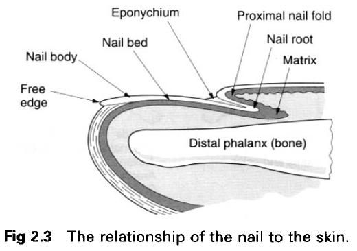
The distal end of the nail is free, while the
proximal covered part constitutes the nail root. There is an abundant supply of
sensory nerve endings and blood vessels to the nail bed. The nails grow at
approximately 1mm per week, being faster in summer than in winter.
Hairs
Hairs are widely distributed over the body
surface, notable exceptions being the palm of the hand and the sole of the
foot. Hairs vary as to their thickness and length. Most of them are extremely
fine so that the skin may appear hairless. There is a marked sexual difference
in the distribution of coarse hair, particularly on the face and trunk, and in
its loss from the scalp. This coarse hair tends to become more prominent after
puberty, particularly in the axilla, over the pubes, and on the face in males.
Except for the eyelashes, all hairs emerge
obliquely from the skin surface, with the hairs in any one region doing so in
the same direction. The part of the hair which projects from the skin surface
is the shaft, with that part under the skin being the root, which is ensheathed
in a sleeve of epidermis known as the follicle
extending into the subcutaneous tissue. The shaft appears circular in
cross-section. Throughout most of its length the hair consists of the
keratinized remains of cells. Hair colour is due to pigment in the hair
cells(melanin and a subtle red pigment), and to air within the shaft of the
hair. The hair of the head has a life of between 2 and 4 years, but that of the
eyelashes is only 3 to 5 months. All hairs are intermittently shed and
replaced.
In the growing hair, the deepest part of the
hair follicle expands to form a cap, known as the bulb of the hair, which
almost completely surrounds some loose, vascular connective tissue, known as
the papilla. The cells of the follicle around the papilla proliferate to form
the various layers of the hair. In the resting hair follicle the bulb and
papilla shrink, with the deepest part of the follicle being irregular in shape.
Associated with each hair are one or more
sebaceous glands, which lie in the angle between the slanting hair follicle and
the skin surface with their ducts opening into the neck of the follicle.
Bundles of smooth muscle fibres(the arrector pullorum) attach to the sheath of
the hair follicle, deep to the sebaceous gland, and pass to the papillary
layers of the dermis on the side towards which the hair slopes(picture b).
Contraction of the muscle causes hair to stand away from the skin, elevating the
skin around the opening of the hair follicles, thereby producing “goose flesh”.
This action also compresses the sebaceous glands causing them to empty their
secretions onto the skin surface. Elevation of the hairs traps a layer of air
against the skin surface in an attempt to produce an insulating layer to reduce
heat loss, while the sebaceous secretions are important in “water-proofing” the
skin surface and in aiding the absorption of fat-soluble substances through the
skin.


0 коментара:
Постави коментар