


The clavicle is a subcutaneous bone running
horizontally from the sternum to the acromion. It acts as a strut holding the
scapula laterally, thus enabling the arm to be clear of the trunk – an
essential feature in primates. The scapula and clavicle together form the
pectoral or shoulder girdle,
transmitting the weight of the upper limb to the axial skeleton and
facilitating a wide range of movement of the upper limb.
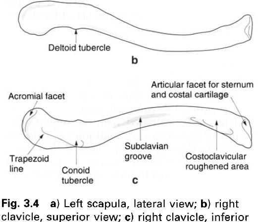
The medial two-thirds of the clavicle is convex
forwards and is roughly triangular in cross-section. The lateral third is
concave forwards and flattened from above downwards. The medial convexity
conforms with the curvature of the superior thoracic aperture, the lateral
concavity with the shape of the shoulder.
The lateral(acromial) end of the clavicle is
the most flattened part of the bone and has a small deltoid tubercle on its anterior border. Inferiorly, the rounded conoid tubercle is present at the
posterior edge of the bone, with the rough trapezoid
line(figure c) running forwards and laterally away from it. The conoid
tubercle and trapezoid line give attachment to the conoid and trapezoid parts
of the coracoclavicular ligament binding the clavicle and scapula together.
Laterally is a small oval facet for the acromion process. It is set obliquely
facing downwards and laterally.
The medial(sternal) end of the clavicle is
enlarged and faces downwards and medially. The lower three-quarters is beveled
and articulates with the clavicular notch of the manubrium and the costal
cartilage of the first rib, forming the sternoclavicular joint. The cylindrical
clavicle projects above the shallow notch of the sternum; this can be confirmed
by palpation. The superior quarter of the sternal end is roughened for the
attachment of the intra-articular disc and ligaments. Between the lateral and
medial ends, the superior surface of the clavicle is smooth, while the inferior
surface is marked by a rough subclavian
groove centrally and a large oval roughened area for the costoclavicular ligament medially. The
anterior and posterior borders are roughened by muscle attachments.
The clavicle is often fractured by the direct
violence of a blow, or by indirect forces transmitted up the limb following a
fall on an outstretched arm. The fracture usually occurs at the junction of the
two curvatures, and the resultant fracture deformity is caused by the weight of
the arm pulling the shoulder downwards and medially.
Palpation
In a slender subject the whole length of the
clavicle can often be seen pressing against the skin. Initially, the enlarged
medial end of the clavicle can be palpated with the fingers, and the line of
the sternoclavicular joint can also be identified. Moving laterally, almost the
whole length of the shaft of the clavicle can be gripped between finger and
thumb. At the lateral end, the bulk of deltoid may require deeper pressure;
nevertheless the line of the acromioclavicular joint should be palpable,
particularly from above.
Ossification
The clavicle ossifies in membrane, being the
first bone in the body to begin ossification. Two primary centres appear during
the fifth week in utero. These centres unite and ossification spreads towards
the ends of the bone. A secondary centre appears in the medial end between 14
and 18 years, fusing with the main part of the bone as early as 18 to 20 years
in females and 23 to 25 years in males. An additional centre may appear in the
lateral end at puberty; however it soon fuses with the main bone.


0 коментара:
Постави коментар