


The humerus is the largest of the bones in the
upper limb. It is a typical long bone, having a shaft(body) and two
extremities. Proximally, the humerus articulates with the glenoid fossa of the
scapula forming the shoulder joint, and distally with the radius and ulna
forming the elbow joint.
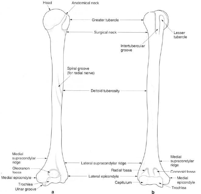
The major feature proximally, is the head of the humerus with its smooth,
rounded articular surface facing upwards, medially and backwards. It is almost
hemispherical, being siderably larger than the socket formed by the glenoid
fossa. The head is joined to the upper end by the anatomical neck, a slightly
constricted region encircling the bone at the articular margin, separating it
from the tubercles.
The greater
tubercle is a prominence on the upper lateral part of the bone, next to the
head. It merges with the shaft below and is marked by three distinct
impressions for muscular attachment. The greater tubercle projects laterally
past the margin of the acromion and is the most lateral bony point at the shoulder.
The smaller lesser
tubercle is a distinct prominence on the anterior aspect below the
anatomical neck. It has a well-marked impression on its medial side for
muscular attachment. Between these two tubercles, and passing onto the shaft of
the humerus, is the deep intertubercular
groove(sulcus). The crests of the greater and lesser tubercles continue
down from the anterior borders of the tubercles to form the lateral and medial
lips of the groove. Between the two lips is the floor of the groove.
Below the head and tubercles, where they join
the shaft, there is a definite constriction. This region is termed the surgical neck because fractures often
occur here, particularly in the elderly.
The shaft of the humerus is almost cylindrical
above, becoming triangular in its lower part with distinct medial and lateral
borders. It presents three borders and surfaces, although the borders are
frequently rounded and indistinct. They are described as anterior, medial and
lateral. Between the three borders are the three surfaces of the shaft. The
intertubercular groove is continuous with the anteromedial surface, the medial
border beginning as the crest of the lesser tubercle and ending by curving
towards the medial epicondyle. The smooth anterolateral surface is marked about
its middle by the deltoid tuberosity.
The posterior surface is crossed obliquely from superomedial to inferolateral
by the spinal groove(radial groove). It reaches the lateral border below the
deltoid tuberosity, but is often poorly marked.
The lower end of the humerus is expanded
laterally, flattened anteroposteriorly, and bent slightly forwards. It presents
two articular surfaces separated by a ridge. The lateral articular surface, the
capitulum, is situated on the
anteroinferior aspect and is a rounded, convex surface, being less than a
hemisphere in size. The capitulum articulates with the radius, making its
greatest contact with the radius when the elbow is fully flexed.
Medial to the capitulum is the trochlea, the articular surface for the
ulna. The trochlea is a grooved surface rather like a pulley, the medial edge
projecting further distally and anteriorly than the lateral. This causes the
ulna also to project laterally and results in a carrying angle between the
humerus and ulna(figure b).
On the medial side of the trochlea is the large
medial epicondyle. Its posterior
surface is smooth and has a shallow groove for the ulnar nerve. The sharp medial supracondylar ridge, comprising
the lower third of the medial border, runs upwards onto the shaft. On the
lateral side of the capitulum is the lateral
epicondyle with the lateral
supracondylar ridge, comprising the lower third of the lateral border,
running upwards onto the shaft.
Just above the articular surfaces, the lower
end of the humerus presents three fossae for the bony processes of the radius
and ulna. Situated posteriorly is the deep olecranon
fossa which receives the olecranon process of the ulna when the elbow is
extended. Anteriorly, there are two fossae: the lateral radial and medial coronoid
fossae, which receive the head of the radius and coronoid process of the ulna
respectively on full flexion of the elbow. Many of the bony features can be
seen in the figure below.
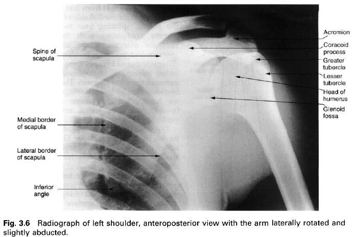
Palpation
At the upper end of the humerus the most lateral
bony point at the shoulder is the greater tubercle, whose quadrilateral
superior, anterior and posterior surfaces can be felt. Further differentiation
can be made by palpating the lateral margin of the acromion and then running
the fingers off its edge onto the greater tubercle. The rounded lesser tubercle
can be left through deltoid, and is just lateral to the hip of the coracoid
process. To the lateral side of the lesser tubercle the impression of the
intertubercular sulcus can usually be felt. The shaft of the humerus is covered
with thick muscle, but can be palpated on its medial and lateral sides. At the
lower end, the prominent medial epicondyle is the most obvious bony landmark.
The ulnar nerve can be rolled in the groove behind it(the “funny bone”).
Running upwards from the medial epicondyle the sharp medial supracondylar ridge
can be palpated. The lateral epicondyle can be palpated at the base of a dimple
on the lateral aspect of the elbow, as can the lateral supracondylar ridge
running upwards from it. Posteriorly, the olecranon fossa can be felt through
the triceps tendon, if the relaxed elbow is flexed.
Ossification
A primary ossification centre appears in the
shaft in the eighth week in utero and
spreads until, at birth, only the ends are cartilaginous. Secondary centres
appear in the head early in the first year, in the greater tubercle at about 3
years and in the lesser tubercle at about 5 years. These fuse to form a single
cap of bone between the ages of 6 and 8 years, finally fusing with the shaft
between 18 and 20 in females and 20 and 22 years in males.
At the lower end of the humerus, secondary
centres appear for the capitulum during the second year, for the trochlea
between 9 and 10 years, and for the lateral epicondyle between 12 and 14 years.
These join together at about 14 years, fusing with the shaft at 15 years in
females and 18 years in males. A separate centre for the medial epicondyle
appears between 15 and 18 years with a spicule of bone projecting down from the
shaft medial to the trochlea. This latter ossification centre lies entirely
outside the joint capsule. Most of the growth in length of the humerus occurs
at its upper end.


0 коментара:
Постави коментар