


Skeletal muscle
Skeletal muscle constitutes over one-third of
the total human body mass. It consists of non-branching striated muscle fibers,
bound together by loose areolar tissue. Muscles have various forms; some are
flat and sheet-like, some are short and thick, while others are long and
slender. The length of a muscle, exclusive of tendons, is closely related to
the distance through which it is required to contract. Experiment has shown
that muscle fibres have the ability to shorten to almost half their resting
length. Consequently, the arrangement of fibres within a muscle determines how
much it can shorten when it contracts. Irrespective of muscle fibre
arrangement, it has to be remembered that all movement is brought about by
muscle shortening, with the consequent action of pulling across joints changing
the relative positions of the bones involved.
Muscle forms
The arrangement of the individual fibres within
a muscle can be in one of two ways only; either parallel or oblique to the line
of pull of the whole muscle. When the fibres are parallel to the line of pull
they are grouped in a discrete bundle giving a fusiform muscle(figure a,
example biceps brachii) or spread as a broad, thin sheet(figure b, example
external oblique of the abdomen). When contraction occurs it does so through
the maximal distance allowed by the length of the muscle fibres. However, it is
of limited power.
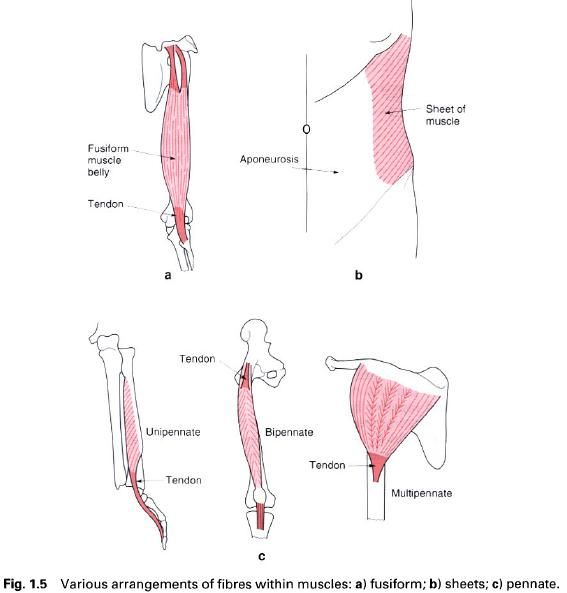
Muscles whose fibres are oblique to the line of
pull cannot shorten to the same degree, but because of the increased number of
fibres packed into the same unit area they are much more powerful. Such
arrangements of fibres are known as pennate,
of which there are three main patterns(figure c). In unipennate muscles the fibres attach to one side of the tendon
only(e.g. flexor pollicis longus). Bipennate
muscles have a central septum with the muscle fibres attaching to both
sides of the septum and to its continuous central tendon(e.g. rectus femoris).
Finally, some muscles possess several intermediate septa, each of which has
associated with it a bipennate arrangement of fibres. The whole is known as a multipennate muscle(e.g. deltoid).
Muscle structure
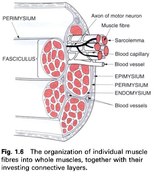
Muscle consists of many individual fibres, each
being a long, cylindrical, multinucleated cell of varying length and width.
Each fibre has a delicate connective tissue covering(endomysium), separating it from its neighbours, yet connecting them
together. Bundles of parallel fibres(fasciculi)
are bound together by a more dense connective tissue covering(perimysium). It is groups of fasciculi
which are bound together to form whole muscles, and are enclosed in a fibrous
covering(epimysium), which may be thick
and strong or thin and relatively weak.
Muscle attachments
The attachment of muscle to bone or some other
tissue is always via the connective tissue elements of the muscle. Sometimes
the perimysium and epimysium unite directly with the periosteum of bone or with
joint capsules. Where this connective tissue element cannot readily be seen,
the muscle has a fleshy attachment and leaves no mark on the bone, although the
area is often flattened or depressed. In many instances the connective tissue
elements of the muscle fuse together to form a tendon, consisting of bundles of
collagen fibres. There is, however, no direct continuity between the fibres of
the muscle and those of the tendon. Tendons can take various forms, all of
which are generally strong. They can be round cords, flattened bands or thin
sheets, the latter being an aponeurosis.
Attachments of tendon to bone nearly always leave a smooth mark; it is only
when the attachment is by a mixture of fleshy and tendinous fibres, or when the
attachment is via a long aponeurosis, that the bone surface is roughened.
Where a muscle or tendon passes over or around
the edge of a bone it is usually separated from the bone by a bursa, which serves to reduce friction
during movement. Bursae are sac-like dilatations which may communicate directly
with an adjacent joint cavity or exist independently, and contain a fluid
similar to synovial fluid.
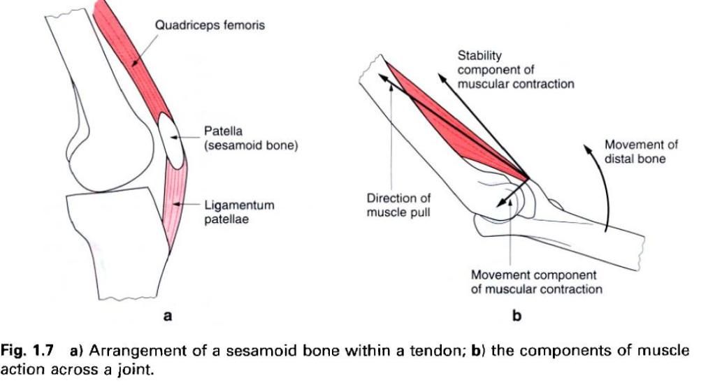
When a tendon is subjected to friction it may
develop a sesamoid bone. Once formed
thse have the general effect of increasing the lever arm of the muscle, and act as pulleys enabling a slight
change in the direction of pull of the muscle, e.g. the patella and the
quadriceps tendon(figure a).
Because each end of a muscle attaches to
different bones, observation of its principal action led to the designation of
tone end being the origin and the other the insertion; the insertion being to
the bone which showed the freest movement. Such a designation is however
misleading, since the muscle can cause either of the two attachments to move
relatively freely. The term attachment
is therefore the preferred one to use.
Muscle action
When stimulated, a muscle contracts so as to
bring its two ends closer together. If this is allowed to happen the muscle
length obviously changes, although the tension generated remains more or less
constant; such contractions are termed isotonic.
If however, the length of the muscle remains unaltered(due to some externally
applied force) then the tension it develops usually increases in an attempt to
overcome the resistance; such contractions are termed isometric.
Isotonic contractions can be of two types, concentric, in which the muscle
shortens, or eccentric, in which the
muscle lengthens. Eccentric contraction occurs when the muscle is being used to
control the movement of a body segment against an applied force.
When a muscle, or group of muscles, contracts
to produce a specific movement, it is termed a prime mover. Muscles which directly oppose this action are called antagonists. Muscles which prevent
unwanted movements associated with the action of the prime movers are known as synergists.
In all actions, part(often the larger) of the
muscle activity is directed across the joint, thereby stabilizing it by pulling
the two articular surfaces together.
When testing the action of a muscle to
determine whether it is weakened or paralysed, the subject is usually asked to
perform the principal action of the muscle against resistance. This may be
insufficient to confirm the integrity of the muscle. The only infallible guide
is to palpate over the muscle belly or its tendons to determine whether it is
contracting during the manoeuvre.


0 коментара:
Постави коментар