


The skin consists of a superficial layer of
ectodermal origin known as the epidermis,
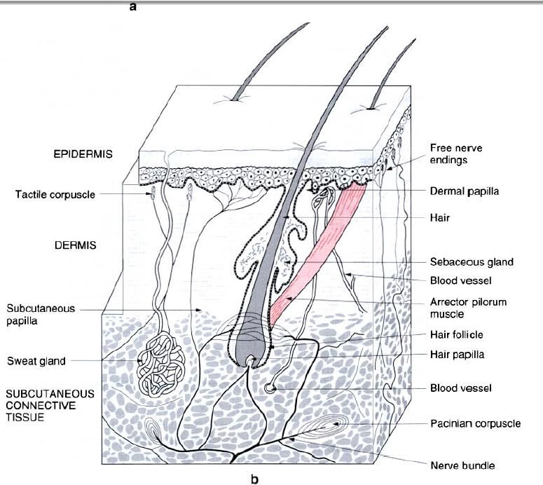
and a deeper mesodermal – derived layer known as the dermis(picture a).
Epidermis
The epidermis is a layer of stratified squamous
epithelium of varying thickness(0.3 – 1.0 mm), being composed of many layers of
cells. The deeper cells are living and actively proliferating, with the cells
produced gradually passing toward the surface. As they do so they become
cornified(keratinized). They are
ultimately shed as the skin rubs against the clothing and other surfaces. The
epidermis is avascular but is penetrated by sensory nerve endings. Its deep
surface is firmly locked to the underlying dermis by projections into it known
as epidermal pegs, with the reciprocal
projections from the dermis being known as dermal
papillae(figure a).

It is usually convenient to consider the
epidermis as being divided into a number of layers, particularly in the
so-called thick skin of the palm or sole of the foot. These layers are from
within outwards known as the stratum
basale, stratum spinosum, stratum granulosum, stratum lucidum and stratum
corneum(figure a).
The stratum basale consists of a single layer
of cells adjacent to the dermis. It is in this layer, as well as in the stratum
spinosum that new cells are produced to replace those lost from the surface.
The stratum spinosum itself consists of several layers of irregularly shaped
cells, which become flattened as they approach the stratum granulosum. The
stratum basale and stratum spinosum together are often reffered to as the
germinal zone, because of their role in new cell production.
Collectively, the remaining layers of the
epidermis(granulosum, lucidium and corneum) are often referred to as the horny
layer. In the stratum granulosum the cells become increasingly flattened and
the process of keratinization begins. The cells in this layer are in the
process of dying. A relatively thin transparent layer(the stratum lucidum) lies
between the granulosum and the superficial stratum corneum. It is this latter
layer from which the cells are shed, and also which is mainly responsible for
the thickness of the skin.
The epidermal
melanocytes, which are responsible for the pigmentation of the skin, lie
within the deepest layers of the epidermis.
Dermis
The dermis is the deeper interlacing feltwork
of collagen and elastic fibres, which generally comprises the greater part of
total skin thickness. It can be divided into a superficial finely – textured papillary layer, which, although clearly
separated from it, interdigitates with the epidermis, and a deeper coarser
reticular layer, which gradually blends into the underlying subcutaneous
connective tissue.
The projecting dermal papillae usually contain
capillary networks which bring the blood into close association with the
epidermis(figure a). The ability to open up or close down these networks is
responsible for the regulation of heat loss through the skin, as well as
causing the individual to blush in moments of embarrassment. Some of the
papillae contain tactile receptors, these obviously being more numerous in
regions of high tactile sensitivity(e.g. fingers, lips) and less so in other
regions(e.g. back).
The reticular layer of dermis consists of a
dense mass of interweaving collagen and elastic connective tissue fibres. It is
this layer which gives the skin its toughness and strength. The tissue fibres
run in all directions, but are generally tangential to the surface. However,
there is a predominant orientation of fibre bundles, with respect to the skin
surface, which varies in different regions of the body. It is this orientation
which gives rise to the cleavage lines of the skin.
The dermis contains the numerous blood vessels and lymphatic
channels, nerves and sensory nerve endings as well as a small amount of fat. In addition to these it also
contains hair follicles, sweat and sebaceous glands, and smooth
muscle(arrectores pili). The deep surface of the dermis is invaginated by
projections of subcutaneous connective tissue, which serve partly for the
entrance of the nerves and blood vessels into the skin(figure b).
Subcutaneous connective tissue
This is a layer of loosely arranged connective
tissue containing fat and some
elastic fibres. The amount of subcutaneous fat varies in different parts of the
body, being completely absent in only a few regions(eyelid, scrotum, penis,
nipple and areola). The distribution of subcutaneous fat also differs between
men and women, constituting a secondary sexual characteristic in women, e.g.
the breast as well as the rounded contour of the hips. The subcutaneous
connective tissue contains blood and lymph vessels, the roots of hair
follicles, the secretory parts of sweat glands, cutaneous nerves, and sensory
endings(particularly Pacinian(pressure)
corpuscles) – figure b.
In the subcutaneous tissue overlying joints,
subcutaneous bursae exist, which contain a small amount of fluid, and thereby
facilitate movement of the skin in these regions.


0 коментара:
Постави коментар