


Muscle fiber types
Not all muscle fibers are alike. A single
skeletal muscle contains fibers having different speeds of shortening and
strength; slow-twitch, or type I,
fibers, and fast-twitch, or type II,
fibers. Slow-twitch fibers take
approximately 110ms to reach peak tension when stimulated. Fast-twitch fibers, on the other hand, can reach peak tension in
about 50ms. While the terms “slow twitch” and “fast twitch” continue to be
used, scientists now preffer to use terminology tupe I and type II, as is the
case here.
Although only one form of type I fiber has been
identified, type II fibers can be further classified. The two major forms of
type II fibers are fast-twitch type a(type IIa) and fast-twitch type x(IIx).
Type IIx fibers in humans are approximately the equivalent of type IIb fibers
in animals. If you look under the electronic microscope, type I fibers will be
stained as black; type IIa fibers appear to be white, and type IIx fibers
appear to be gray. Although not visible under the microscope, there is also
third subtype of fast-twitch fibers that has been identified, IIc fibers.
The differences among the type IIa, type IIx,
and type IIc fibers are not fully understood, but type IIa fibers are believed
to be the most frequently recruited. Only type I fibers are recruited more
frequently than type IIa fibers. Type IIc fibers are the least often used. On
the average, most muscles are composed of roughly 50% type I fibers and 25%
type IIa fibers. The remaining 25% are mostly type IIx, with type IIc fibers
making only 1-3% of the muscle. Cause knowledge about type IIc is limited,
there will be no further discussion about that thema. The exact percentage of
each fiber types varies greatly in various muscles and among individuals, so
the numbers listed here are only average values. This extreme variation is most
evident in the athletes, as we will see later when comparison table appears.
Characteristics of type I and type II fibers
Different muscle fiber types play different
roles in physical activity. This is largely due to differences in their characteristics.
ATPase
The type I and type II fibers differ in their
speed of contraction. This difference results primarily from different forms of
myosin ATPase. Recall that myosin ATPase in the enzyme that splits ATP to
release energy to drive contraction. Type I fibers have a slow form of myosin
ATPase, whereas type II fibers have a fast form. In response to neural
stimulation, ATP is split more rapidly in type II fibers than in type I fibers.
As a result, cross-bridges cycle more rapidly in type II fibers.
One of the methods used to classify muscle
fibers is a chemical staining procedure applied to a thin slice of tissue. This
staining technique measures the ATPase activity in the fibers. Thus, the type
I, type IIa, and type IIx fibers stain differently. This technique makes it
appear that each muscle fiber has only one type of ATPase, but fibers can have
a mixture of ATPase types. Some have a predominance of type I-ATPase, but
others have mostly type II-ATPase. Their appearance in a stained slide preparation
should be viewed as a continuum rather than as absolutely distinct types.
A newer method for identifying fiber types is
to chemically separate the different types of myosin molecules(isoforms) by
using a process called gel electrophoresis. The isoforms are separated and
stained to show the bands of protein that characterize type I, type II and type
IIx fibers. Although our discussion here cathegorizes fiber type simply as slow
twitch(type I) and fast twitch(type IIa and type IIx), scientists have further
subdivided these fiber types. The use of electrophoresis has led to the
detection of myosin hybrids or bigers that possess two or more forms of myosin.
With this method of analysis, the fibers are classified as I; Ic(I/IIa);
IIc(IIa/I); IIa; IIax; IIxa; and IIx.
The table below shows the characteristics of
different muscle fiber types. The table also includes alternative names that
are used in other classification systems to refer to the various muscle fiber
types.
Table
1. Classification of muscle fiber types
|
Fiber
classification
|
|||||
|
System
1
|
Type I
|
Type IIa
|
Type IIx
|
||
|
System
2
|
Slow twitch(ST)
|
Fast twitch a(FTa)
|
Fast twitch x (FTx)
|
||
|
System
3
|
Slow oxidative(SO)
|
Fast oxidative/glycolitic(FGO)
|
Fast glycolytic(FG)
|
||
|
Characteristics
of fiber types
|
|||||
|
Oxidative
capacity
|
High
|
Moderately High
|
Low
|
||
|
Glycolitic
capacity
|
Low
|
High
|
Highest
|
||
|
Contractile
speed
|
Slow
|
Fast
|
Fast
|
||
|
Fatique
resistance
|
High
|
Moderate
|
Low
|
||
|
Motor
unit strength
|
Low
|
High
|
High
|
||
Sarcoplasmic
reticulum
Type II fibers have a more highly developed SR
than do type I fibers. Thus, type II fibers are more adept at delivering
calcium into the muscle cell when stimulated. This ability is thought to
contribute to the faster speed of contraction(Vo) of type II fibers.
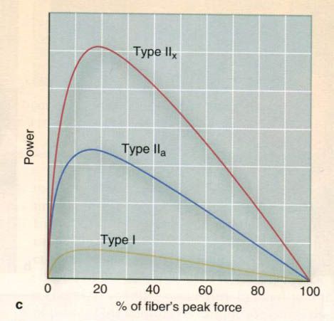
On average, human type II fibers have Vo a that is five to six times
faster than of type I fibers. Although the amount of force(Po)
generated by type II and type I fibers having the same diameter is about the
same, the calculated power(μN x fiber length-1 x s-1) of
a type II fiber is three to five times greater than that of type I fiber
because of a faster shortening velocity. This may explain in part why
individuals who have a predominance of type II fibers in their leg muscles tend
to be better sprinters than individuals who have a high percentage of type I
fibers.
Motor units
Recall that a
motor unit is composed of a single alpha-motor neuron and muscle fibers it
innervates. The alpha motor neuron appears to determine whether the fibers are
type I or type II. The alpha motor neuron in a type I motor unit has smaller
cell body and typically innervates a cluster of less or equal 300 muscle
fibers. This difference in the size of motor units means that when a single
type I alpha-motor neuron stimulates its fibers, far fewer muscle fibers
contract than when a single type II alpha-motor neuron stimulates its fibers.
Consequently, type II muscle fibers reach peak tension faster and collectively
generate more force than type I fibers.
Distribution of fiber types
As mentioned
earlier, the percentages of type I and type II fibers are not the same in all
the muscles of the body. Generally, a person’s arm and leg muscles have similar
fiber compositions. Studies have shown that people with a predominance of type
I fibers in their leg muscles will likely have a high percentage of type I
fibers in their arm muscles as well. A similar relationship exists type II
fibers. There are some exceptions, however. The soleus muscle(beneath the
gastrocnemius in the calf), for example, is composed of a very high percentage of
type I fibers in everyone.
Fiber type and exercise
Because of these
differences in type I and type II fibers, one might expect that these fiber
types would also have different functions when people are physically active.
Indeed, this is the case.
Type I fibers
In general, type
I muscle fibers have a high level of aerobic endurance. Aerobic means “in the
presence of oxygen”, so oxidation is an aerobic process. Type I fibers are very
efficient at producing ATP from the oxidation of carbohydrate and fat.
Recall that ATP
is required to provide the energy needed for muscle fiber contraction and
relaxation. As long as oxidation occurs, type I fibers continue producing ATP,
allowing the fibers to remain active. The ability to maintain muscular activity
for a prolonged period is known as muscular endurance, so type I fibers have
high aerobic endurance. Because of this, they are recruited most often during
low-intensity endurance events(e.g. marathon running) and during most daily
activities for which the muscle force requirements are low(e.g. walking).
Type II fibers
Type II muscle
fibers, on the other hand, have relatively poor aerobic endurance when compared
to type I fibers. They are better suited to perform anaerobically(without
oxygen). This means that in the absence of adequate oxygen, ATP is formed
through anaerobic pathways, not oxidative pathways.
Type IIa motor
units generate considerably more force than do type I motor units, but type Iia
motor units also fatique more easily because of their limited endurance. Thus,
type Iia fibers appear to be the primary fiber type used during shorter,
higher-intensity endurance events, such as the mile run or the 400m swim.
Although the
significance of the type IIx fibers is not fully understood, they apparently are
not easily activated by the nervous system. Thus they are used rather
infrequently in normal, low-intensity activity but are predominantly used in
highly explosive events such as the 100m dash and the 50m sprint swim.
Characteristics of the various fiber types are set in the table below.
Table 1.2. Structural and functional
characteristics of muscle fiber types
|
|
Fiber type
|
||
|
Characteristic
|
Type I
|
Type IIa
|
Type IIb
|
|
Fibers per motor neuron
|
≤ 300
|
≥300
|
≥300
|
|
Motor neuron size
|
smaller
|
larger
|
larger
|
|
Nerve conduction velocity
|
slower
|
faster
|
faster
|
|
Contraction speed(ms)
|
110
|
50
|
50
|
|
Type of myosin ATPase
|
slow
|
fast
|
fast
|
|
Sarcoplasmic reticulum development
|
low
|
high
|
high
|
Determination of fiber
type
The
characteristics of muscle fibers appear to be determined early in life, perhaps
within the first few years. Studies with identical twins have shown that muscle
fiber type, for the most part, is genetically determined, changing little from
childhood to middle age. These studies reveal that identical twins have nearly
identical fiber types, whereas fraternal twins differ in their fiber type
profiles. The genes we inherit from our parents likely determine which
alpha-motor neurons innervate our individual muscle fibers. After innervation
is established, muscle fibers differentiate(become specialized) according to
the type of alpha-motor neuron that stimulates them. Some recent evidence,
however, suggests that endurance training, strength training, and muscular
inactivity may cause a shift in the myosin isoforms. Consequently, training may
induce a small change, perhaps less than 10%, in the percentage of type I and
type II fibers. Further, both endurance and resistance training have been shown
to reduce the percentage of type IIx fibers while increasing the fraction of
type Iia fibers.
Studies of older
men and women have shown that aging may alter the distribution of type I and
type II fibers. As we grow older, muscles tend to lose type II motor units,
which increases the percentage of type I fibers.
Muscle fiber recruitment
When an
alpha-motor neuron carries an action potential to the muscle fibers in the
motor unit, all fibers in the unit develop force. Activating more motor units
produces more force. When little force is needed, only a few motor units are
stimulated to act. Recall from our earlier discussion that type Iia and type
Iix motor units contain more muscle fibers than type I motor units do. Skeletal
muscle contraction involves a progressive recruitment of type I and then type
II motor units, depending on the requirements of the activity being performed.
As the intensity of the activity increases, the number of fibers recruited
increases in the following order, in an additive manner: type I à type IIa à type IIx.
Orderly recruitment of
muscle fibers and the size principle
Most researchers
agree that motor units are generally activated on the basis of a fixed order of
fiber recruitment. This is known as the principle of orderly recruitment,
in which the motor units within a given muscle appear to be ranked. Let’s use
the biceps brachii as an example: assume a total of 200 motor units, which are
ranked on a scale from 1 to 200. For an extremely fine muscle contraction
requiring very little force production, the motor unit ranked number 1 would be
recruited. As the requirements for force production increase, numbers 2,3,4;
and so on would be recruited, up to a maximal muscle contraction that would
activate most, if not all, of the motor units. For the production of a given
force, the same motor units are usually recruited each time and in same order.
A mechanism that
may partially explain the principle of orderly recruitment is the size
principle, which states that the order of recruitment of motor units is
directly related to their motor neuron size. Motor units with smaller motor
neurons will be recruited first. Because the type I motor units have smaller
motor neurons, they are the first units recruited in graded movement(going from
very low to very high rates of force production). The type II motor units then
are recruited as the force needed to perform the movement increases. It is
unclear at this time how the size principle relates to complex athletic
movements.
During events
that last several hours, exercise is performed at a submaximal pace, and the
tension in the muscles is relatively low. As a result, the nervous system tends
to recruit those muscle fibers best adapted to endurance activity: the type I
and some type IIa fibers. As the exercise continues, these fibers become
depleted of their primary fuel supply(glycogen), and the nervous system must
recruit more type IIa fibers to maintain muscle tension. Finally, when the type
I and type IIa fibers become exhausted, the type IIx fibers may be recruited to
continue exercising.
This may explain
why fatique seems to come in stages during events such as the marathon, a 42km
run. It also may explain why it takes great conscious effort to maintain a
given pace near the finish of the event. This conscious effort results in the
activation of muscle fibers that are not easily recruited. Such information is
of practical importance to our understanding of the specific requirements of
training and performance.
Use of muscles
Types of muscle
contraction
Muscle movement
generally can be cathegorized into three types of contractions – concentric,
static and eccentric. In many activities, such as running and jumping, all
three types of contraction may occur in the execution of a smooth, coordinated
movement. For the sake of clarity, though, we will examine each type separately.
A muscle’s
principal action, shortening, is reffered to as a concentric contraction,
the most familiar type of contraction. To understand muscle shortening, recall
our earlier discussion of how the thin and thick filaments slide across each other.
In a concentric contraction, the thin filaments are pulled toward the center of
the sarcomere. Because joint movement is produced, concentric contractions are
considered dynamic contractions.
Muscles can also
act without moving. When this happens, the muscle generates force, but its
length remains static(unchanged). This is called a static, or isometric,
muscle contraction, because the joint angle does not change. A
static contraction occurs, for example, when one tries to lift an object that
is heavier than the force generated by the muscle, or when one supports the
weight of an object by holding it steady with the elbow flexed. In both cases,
the person feels the muscles tense, but there is no joint movement. In a static
contraction, the myosin cross-bridges form and are recycled, producing force,
but the external force is too great for the thin filaments to be moved. They
remain in their normal position, so shortening can’t occur. If enough motor
units can be recruited to produce sufficient force to overcome the resistance,
a static contraction can become a dynamic one.
Muscles can
exert force even while lengthening. This movement is an eccentric contraction.
Because joint movement occurs, this is also a dynamic contraction. An example
of an eccentric contraction is the action of the biceps brachii when one
extends the elbow to lower a heavy weight. In this case, the thin filaments are
pulled further away from the center of the sarcomere, essentially stretching
it.
Generation of force
Whenever muscles
contract, whether the contraction is concentric, static or eccentric, the force
developed must be graded to meet the needs of the task or activity. Using golf
as an example, the force needed to tap in a 1m(~40 in.) putt is far less than
that needed to drive the ball 100m (109yd) from the tee to the middle of the
fairway. The amount of muscle force developed is dependent on the number and
type of motor units activated, the frequency of stimulation of each motor unit,
the size of the muscle, the muscle fiber and sarcomere length, and speed of
muscle contraction.
Motor units and muscle size
More force can
be generated when more motor units are activated. Type II motor units generate
more force than type I motor units because a type II motor unit contains more
muscle fibers than a type I motor unit. In a similar manner, larger muscles,
having more muscle fibers, can produce more force than smaller muscles.
Frequency of stimulation of the motor
units: rate coding
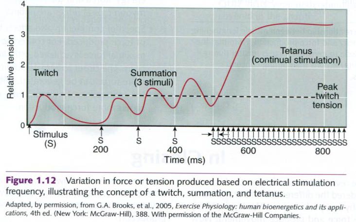
A single motor
unit can exert varying levels of force dependent on the frequency at which it
is stimulated. The smallest contractile response of a muscle fiber or a motor
unit to a single electrical stimulus is termed a twitch. A series of
three stimuli in rapid sequence, prior to complete relaxation from the first
stimulus, can elicit an even greater increase in force or tension. This is
termed summation. Continued stimulation at higher frequencies can lead
to the state of tetanus, resulting in the peak force or tension of the
muscle fiber or motor unit. Rate coding is the term used to describe the
process by which the tension of a given motor unit can vary from that of a
twitch to that of tetanus by increasing the frequency of stimulation of that
motor unit.
Muscle fiber and sarcomere length

There is an
optimal length of each muscle fiber relative to its ability to generate force.
Recall that a given muscle fiber is composed of sarcomeres connected end to end
and that these sarcomeres are composed of both thick and thin filaments. The
optimal sarcomere length is defined as that length where there is optimal
overlap of the thick and thin filaments, thus maximizing cross-bridge
interaction. When a sarcomere is fully stretched(A) or shortened(E), little or
no force can be developed since there is little cross-bridge interaction.
Speed of contraction
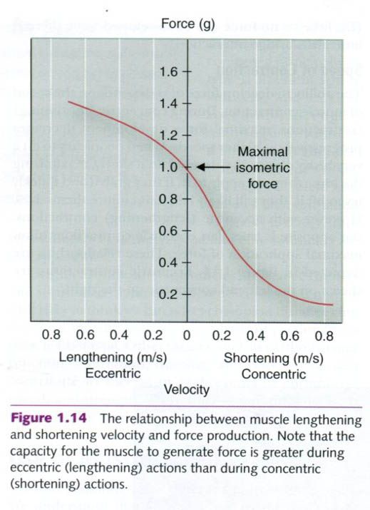
The ability to
develop force also depends on the speed of muscle contraction. During
concentric(shortening) contractions, maximal force development decreases
progressively at higher speeds. When people try to lift a very heavy object,
they tend to do it slowly, maximizing the force they can apply to it. If they
grab it and quickly try to lift it, they will likely fail, if not injure
themselves. However, with eccentric(lengthening) contractions, the opposite is
true. Fast eccentric contractions allow maximal application of force. Eccentric contractions are shown at the left and concentric at the right.
“Physiology of sport and exercise”, fourth
edition; Jack H. Wilmore, David L. Costill, W. Larry Kenney


2 коментара:
Fiber Distribution Unit is essential to build up speed to the servers
Постави коментар