


Neuromuscular junction
Whereas neurons communicate with other neurons
at synapses, an alpha-motor neuron communicates with muscle fibers at a site
known as a neuromuscular junction.
The function of the neuromuscular junction is essentially the same as that of a
synapse. In fact, the proximal part of the neuromuscular junction is the same: It
starts with the axon terminals of the motor neuron, which release
neurotransmitters into the space between the motor nerve and the muscle fiber
in response to an action potential. However, in the neuromuscular junction,
the axon terminals protrude into motor end plates, which are troughlike
segments on the plasmalemma. Picture below shows.
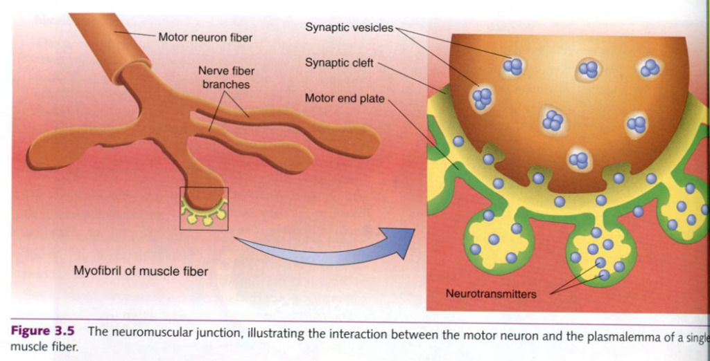
The motor end plate is invaginated(folded to
form cavities). The cavity thus formed is called the synaptic gutter. As with
synapses, the space between the neuron and the muscle fiber is the synaptic cleft.
Neurotransmitters
released from the alpha-motor neuron axon terminals diffuse across the synaptic
cleft and bind to receptors on the muscle fiber’s plasmalemma. This binding
typically causes depolarization by opening sodium ion channels, allowing more
sodium to enter the muscle fiber. As always, if the depolarization reaches the
threshold, an action potential is formed. It spreads across the plasmalemma
into the T-tubules, inititating muscle fiber contraction. As in the neuron, the
plasmalemma, once depolarized, must undergo repolarization. During the period
of repolarization, the sodium gates are closed and the potassium gates are
open; thus, like the neuron, the muscle fiber is unable to respond to any
further stimulation. This period is reffered to as the refractory period. Once
the electrical conditions of the muscle fiber are restored to resting levels,
the fiber can respond to another stimulus. Thus, the refractory period limits
the motor unit’s firing frequency.
Now we know how the impulse is transmitted
between two cells. But to understand what happens once the impulse is
transmitted, we must first examine the chemical signals that accomplish
transmission.
Neurotransmitters
More than 50 neurotransmitters have been
positively identified or are suspected as potential candidates. These can be
cathegorized as either (a) small-molecule, rapid-acting neurotransmitters
or (b) neuropeptide, slow-acting neurotransmitters. The small-molecule,
rapid-acting transmitters, which are responsible for most neural transmissions,
are our main concern.
Acetylholine and norepinephrine are the two
major neurotransmitters involved in regulating our physiological responses to
exercise. Acetylholine is the
primary neurotransmitter for the motor neurons that innervate skeletal muscle
and for most parasympathetic neurons. It is generally an excitatory
neurotransmitter, but it can have inhibitory effects at some parasympathetic
nerve endings, such as in the heart. Norepinephrine
is the neurotransmitter for the most sympathetic neurons, and it too can be
either excitatory or inhibitory, depending on the receptors involved.
Once the neurotransmitters binds to the
post-synaptic receptor, the nerve impulse has been successfully transmitted.
The neurotransmitter is then either degraded by enzymes, actively transported
back into the presynaptic terminals for reuse, or diffused away from the
synapse.


0 коментара:
Постави коментар