


Brachialis
Brachioradialis
Pronator teres
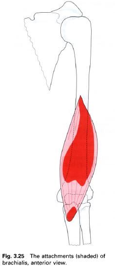
Brachialis
Brachialis lies under cover of biceps brachii in the lower half of the
anterior aspect of the arm. It arises from the distal two-thirds of the anterior
surface of the shaft of the humerus extending outwards onto the
medial and lateral intermuscular septa. The muscle fibers are separated from
the lower part of the lateral intermuscular septum by brachioradialis, with
which it may be partly fused, and extensor carpi radialis longus. The fibres
converge to a thick tendon which forms the floor of the cubital fossa and
attaches to the rough triangular
brachialis impression on the inferior
part of the coronoid process and tuberosity of the ulna. Some deeper fibres of brachialis insert into the capsule of
the elbow joint and serve to pull it away from the moving bones during flexion
and prevent them from becoming trapped.
Nerve
supply
Brachialis is supplied mainly by the musculocutaneous nerve, root value C5
and 6; but also receives a branch from the radial
nerve; again root value C5 and 6, as it runs along the lateral border of
the muscle. However, this latter branch is thought to be almost entirely
sensory.
Action
Brachialis is the main flexor of the elbow
joint.
Functional
activity
Although brachialis flexes the elbow, it is
important in controlling extension produced by gravity. In this situation, the
flexors of the elbow control the movement by an eccentric contraction.
Palpation
When biceps brachii has been identified, brachialis can be felt extending either side
of its belly with the elbow flexed. The tendon can be palpated by applying deep
pressure just above the coronoid process of the ulna.
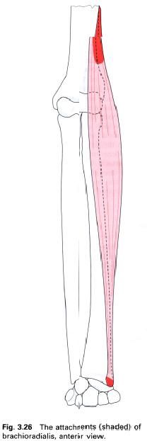
Brachioradialis
Brachioradialis is a superficial muscle found
on the lateral side of the forearm extending almost as far as the wrist. It
forms the lateral border of the cubital fossa, and is covered in its upper part
by brachialis, with which it may be partly fused. The proximal attachment of
brachioradialis is to the upper
two-thirds of the front of the lateral supracondylar ridge of the humerus and the adjacent part of the lateral intermuscular septum. From here,
the fibres run downwards forming a long, narrow, flat tendon in the middle of
the forearm. The tendon is crossed by those of abductor pollicis longus and
extensor pollicis brevis before it attaches to the lateral surface of the radius just above the styloid process.
Nerve
supply
Brachioradialis is supplied by a branch from
the radial nerve, root value C5 and
6, which enters its medial side above the elbow. The skin over the muscle is
also supplied by roots C5 and 6.
Action
Brachioradialis flexes the elbow joint
particularly when the forearm is midway between pronation and supination. It
also helps to return the forearm to this mid position from the extremes of
either pronation or supination; this can be confirmed by palpation.
Functional
activity
Brachioradialis acts primarily to maintain the
integrity of the elbow joint since its fibres run more or less parallel to the radius. It also works eccentrically as
an extensor of the elbow joint in activities such as hammering.
Palpation
With the elbow flexed to 90° and forearm in a
mid-pronated position, the brachioradialis can be felt along the top of the
forearm when the position is maintained against resistance. Using firm
pressure, the tendon can be palpated proximal to the radial styloid process.
The brachioradialis reflex can be elicited by firmly tapping its tendon just
above the wrist.
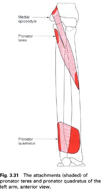
Pronator teres
Pronator teres forms the medial border of the
cubital fossa at the elbow, and as such is the most lateral of the superficial
muscles in the flexor compartment of the forearm. It arises by two heads: the humeral head and the ulnar head.
The humeral head arises from the lower part of the medial supracondylar ridge and adjacent intermuscular septum.
It also comes from the common flexor origin on the medial epicondyle and the covering fascia. The ulnar head
arises from the pronator ridge, which
runs downwards from the medial part of the coronoid
process, to join with the humeral head on its deep surface. Between these
two heads passes the median nerve. The muscle fibres pass downwards and
laterally to attach via a flattened tendon into a roughened oval area on the
middle of the lateral surface of the radius.
Nerve
supply
Pronator teres is supplied by the median nerve, root values C6, 7. The
overlying skin is supplied by roots C6 and T1.
Action
Pronator teres pronates the forearm by
producing an anteromedial swing of the
lower end of the radius across the ulna. This movement of the radius carries the hand with it.
Pronator teres is also a weak flexor of the elbow.
Palpation
The muscle can be palpated running along the
medial border of the cubital fossa between the medial epicondyle of the humerus and the middle of the radius. Pronator teres can be most
easily felt, and occasionally seen, when resisting pronation of the forearm.


0 коментара:
Постави коментар