


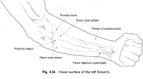
Flexor carpi ulnaris
Flexor carpi radialis
Palmaris longus
Flexor digitorum superficialis
Flexor digitorum profundus
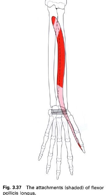
Flexor pollicis longus
Flexor carpi ulnaris
Flexor carpi ulnaris lies along the medial
border of the forearm, being the most medial of the superficial flexor group.
It arises from the humerus and ulna. The humeral head arises from the common flexor origin on the medial epicondyle of the humerus and the adjacent fascia. The
ulnar head arises from the medial border
of the olecranon and, by an
aponeurotic attachment, from the upper two-thirds of the posterior border of
the ulna. Between these two heads
passes the ulnar nerve to gain the medial side of the flexor compartment of the
forearm.
The muscle forms a long tendon about halfway
down the forearm. This attaches to and invests the pisiform. The tendon is prolonged to reach the hook of the hamate and base
of the fifth metacarpal by the pisohamate and pisometacarpal ligaments
respectively. Occasionally, some fibres may be prolonged into abductor digiti
minimi. Lateral to the tendon are the ulnar nerve and vessels.
Nerve
supply
Flexor carpi ulnaris is supplied by several
branches from the ulnar nerve, root
value C7, 8. The skin overlying the muscle is supplied by roots C8 and T1.
Action
In conjunction with flexor carpi radialis, and
to some extent palmaris longus, flexor carpi ulnaris is a flexor of the hand at the wrist. However, when working
with extensor carpi ulnaris, it produces adduction or ulnar deviation of the
hand at the wrist. However, when working with extensor carpi ulnaris, it
produces adduction or ulnar deviation of the hand at the wrist. It also plays an important role in stabilizing
the pisiform during abduction of the little finger, so that abductor digiti
minimi has a firm base from which to work. As with flexor carpi radialis,
flexor carpi ulnaris is an important synergist in extension of the fingers,
preventing unwanted extension of the wrist.
Palpation
The tendon of flexor carpi ulnaris can easily
be identified running proximally from the pisiform where it can be pinched
between the thumb and index finger. In the upper medial part of the forearm,
the muscle belly can be palpated when flexion of the wrist is performed against
resistance.
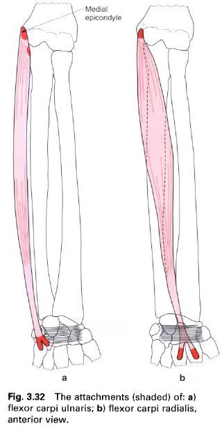
Flexor carpi radialis
Flexor carpi radialis is a fusiform muscle and
is the most lateral of the superficial flexor muscles in the lower half of the
forearm. It arises from the medial
epicondyle of the humerus via the
common flexor tendon and the adjacent
fascia. Halfway down the forearm, the muscle fibres condense to form a long
tendon which passes beneath the flexor retinaculum, where it lies in its own
lateral compartment within the carpal tunnel. Here the tendon is surrounded by
its own synovial sheath as it grooves the trapezium. Distally the tendon
inserts into the palmar surface of the bases of the second and third metacarpals. Its course in the forearm is oblique,
running from medial to lateral and from above downwards. At the wrist, the
tendon lies between the radial vessels laterally and median nerve medially.
Nerve
supply
Flexor carpi radialis is supplied by the median nerve, root value C6, 7. The skin
over the muscle is supplied by roots C6 and T1.
Action
Working with palmaris longus and flexor carpi
ulnaris, flexor carpi radialis acts as a flexor of the wrist. Abduction or
radial deviation of the wrist is produced by the combined action of flexor
carpi radialis and extensors carpi radialis longus and brevis. Because of its
oblique course in the forearm, flexor carpi radialis may aid pronation. It can
also help to flex the elbow. It works in a similar way to flexor carpi ulnaris
in preventing unwanted extension of the wrist when extending the fingers.
Palpation
When the wrist joint is flexed and abducted,
the tendon of flexor carpi radialis can be palpated as the most lateral of the
tendons on the anterior aspect of the wrist, at the level of the radial styloid
process.
Palmaris longus
Palmaris longus is a small vestigial muscle,
being absent in about 10% of the population. Lying centrally among the
superficial flexor muscles of the forearm, palmaris longus arises from the
front of the medial epicondyle of the
humerus via the common flexor origin. The short muscle fibers soon form a long and
slender tendon which passes distally to attach to the superficial surface of
the flexor retinaculum and inserts
into the apex of the palmar aponeurosis. At the wrist, the
tendon lies on top of the median nerve.

Nerve
supply
Palmaris longus is supplied by the median nerve, root value C8. The skin
over the muscle is supplied by roots C7 and T1.
Action
Palmaris longus is a weak flexor of the wrist.
However, because of its attachment to the palmar aponeurosis, it may have some
slight action in flexing the metacarpophalangeal joints and it tightens the
palmar fascia.
Palpation
The tendon of palmaris longus can be identified
just proximal to the wrist, where it is the most central structure when flexion
of the wrist is resisted. The tendon lies on the medial side of that of flexor
carpi radialis.
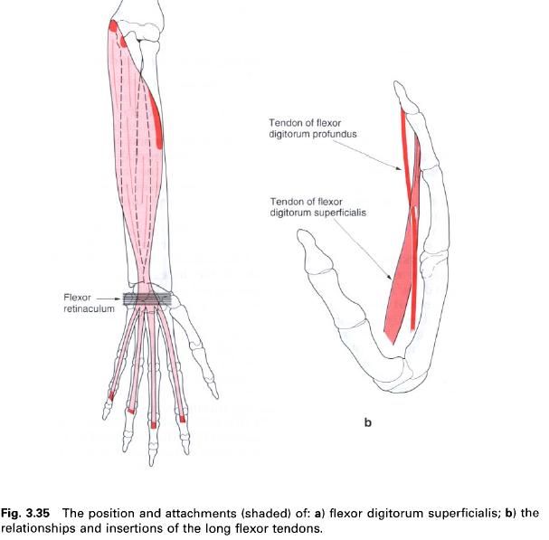
Flexor digitorum superficialis
Flexor digitorum superficialis lies in the
anterior compartment of the forearm deep to pronator teres, palmaris longus and flexors carpi radialis and ulnaris. It is
superficial to flexor digitorum profundus and flexor pollicis longus. This
large muscle has a long, linear origin but may be considered to arise by two
heads. The medial or humeroulnar head arises from: the medial epicondyle via the common
flexor tendon, the anterior part of the ulnar
collateral ligament and the sublime
tubercle at the upper medial part of the coronoid process of the ulna.
The lateral or radial head arises from the upper
two-thirds of the anterior border
of the radius, which runs downwards
and laterally from the radial tuberosity.
About halfway down the forearm, the muscle
narrows to form four separate tendons which pass deep to the flexor retinaculum
where they are arranged in two pairs to enter the hand. The superficial pair pass to the middle and ring fingers,
while the deep pair pass to the index and little fingers. Within the carpal
tunnel the tendons of superficialis are superficial to those of profundus, with
which they share a common synovial sheath. In the palm, the tendons separate
and pass towards their respective fingers, still lying superficial to the
profundus tendons. At the level of the metacarpophalangeal joint, each
superficialis tendon splits longitudinally into two parts. The two halves pass
around the profundus tendon, twisting so that their outer surfaces unite to
form a groove along which the tendon of flexor profundus passes. Prior to
attaching to either side of the palmar
surface of the base of the middle phalanx, the tendon splits again.
This peculiar arrangement of the superficialis
tendons provides a tunnel which allows the profundus tendon to become
superficial. The effect, as fat as superficialis is concerned, is to increase
the lever arm of the tendon at the proximal interphalangeal joint, thereby
enabling a powerful grip of the fingers to be exerted. As well as their main
attachment to the middle phalanx, the tendons of superficialis also provide
attachments for the vincula tendinium which convey blood vessels to the tendon.
Nerve
supply
Flexor digitorum superficialis is supplied by
the median nerve, root value C7, 8,
T1. The skin overlying the muscle and its tendons are supplied by roots C6, 7,
8 and T1.
Action
Flexor digitorum superficialis is primarily a
flexor of the metacarpophalangeal and proximal interphalangeal joints. Because
it crosses the wrist joint, it will also help flexion of the wrist if its
action is continued.
Palpation
Contraction of flexor digitorum superficialis
can be felt by applying deep pressure through the superficial flexor muscles in
the upper part of the forearm whilst the fingers are flexed. The tendons can be
palpated in a similar manner proximal to the flexor retinaculum. The muscle can
be tested specifically by asking the subject to flex the proximal
interphalangeal joint without flexing the distal interphalangeal joint.
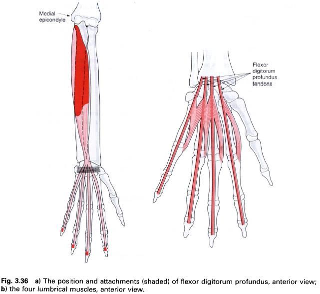
Flexor digitorum profundus
Flexor digitorum profundus lies deep to
superficialis on the medial side of the forearm. It arises from the medial side
of the coronoid process of the ulna, the upper three-quarters of the anterior
and medial surfaces of the ulna, and from the medial, middle-third of the anterior surface of the adjacent interosseus membrane. It also arises from the aponeurosis attaching
flexor carpi ulnaris to the posterior
border of the ulna.
The part of the muscle arising from the
interosseus membrane forms a separate tendon about halfway down the forearm
which passes to the index finger. The remaining tendons are not usually formed
until just above the flexor retinaculum. The separate tendons, however, pass below
the flexor retinaculum where they lie side-by-side, deep to those of
superficialis, but within the same synovial sheath. In the palm, the four
tendons pass to their respective fingers. At first, they travel deep to
superficialis, but then pass superficially as they go through the tunnel formed
by the superficialis tendon at the level of the metacarpophalangeal joint.
The tendon of profundus eventually inserts into
the base of the palmar surface of the distal
phalanx having passed through a fibro-osseous tunnel. Like the tendons of
flexor digitorum superficialis, the profundus tendons are provided with vincula
tendinium.
Nerve
supply
Flexor digitorum profundus has a dual nerve
supply. The lateral part of the muscle, which gives off tendons to the index
and middle fingers, is supplied by the anterior
interosseus branch of the median
nerve, root value C7, 8, T1. The medial part of the muscle, which gives off
tendons to the ring and little fingers, is supplied by the ulnar nerve, root value C8, T1. The skin over the muscle is
supplied by roots C7, 8, T1.
Action
The primary action of flexor digitorum
profundus is flexion of the distal interphalangeal joint. However, because it
crosses several other joints during its course, it also aids in flexion of the proximal
interphalangeal, metacarpophalangeal and wrist joints.
Palpation
The muscular part of profundus can be palpated
immediately medial to the posterior border of the ulna, where its concentration can be felt as the fingers are fully
flexed from a position of extension.
Flexor pollicis longus
Flexor pollicis longus lies on the lateral side
of flexor digitorum profundus. It arises from the anterior surface of the radius
between the radial tuberosity above and pronator quadratus below, and the adjacent
anterior surface of the interosseus membrane. Occasionally, the
muscle also arises by a small slip from the medial border of the coronoid
process of the ulna. The fibres pass almost to the wrist before a single tendon
is formed.
The tendon of flexor pollicis longus passes
below the flexor retinaculum, in its own synovial sheath, to insert into the
palmar surface of the base of the distal
phalanx of the thumb.
Nerve
supply
Flexor pollicis longus is supplied by the anterior interosseus branch of the median nerve, root value C8, T1.
Action
Flexor pollicis longus is the only flexor of
the interphalangeal joint of the thumb, and is thus vital for all gripping
activities of the hand. It also
flexes the metacarpophalangeal joint of the thumb and the wrist joint.
Palpation
When only the thumb is flexed, the contraction
of flexor pollicis longus can be felt in the lower third of the forearm,
immediately lateral to the superficial flexor tendons.


0 коментара:
Постави коментар