


The pelvic girdle comprises three separate
bones, the two innominate bones and
the sacrum, the ring of bone so
formed uniting the trunk and the lower limbs. The innominate is a large and
irregular bone with two expanded triangular blades twisted 90° with respect to
each other in the region of the acetabulum.
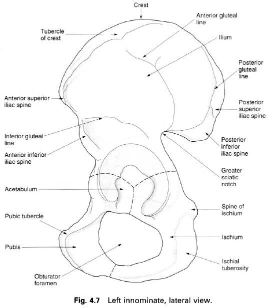
Each triangular blade is also twisted within itself. The innominate bone is
formed from three separate bones, the ilium,
ischium and pubis, which come
together and fuse in the region of the acetabulum so that in the adult the
innominate appears as a single bone. The sacrum is made up of five fused
vertebrae and is roughly triangular. The coccyx,
which is the remnant of our tail, consists of four fused coccygeal vertebrae.
Each innominate bone articulates with the sacrum posteriorly by joints which
are synovial(anteriorly) and fibrous(posteriorly). The innominate bones also
articulate with each other anteriorly at the pubic symphysis, by a secondary
cartilaginous joint.
The pelvis has several functions:
- it supports and protects the pelvic viscera;
- it supports the weight of the body transmitted through the vertebrae, thence though the sacrum, across the sacroiliac joints to the innominate bones and then to the femora in the standing position, or to the ischial tuberosites when sitting;
- during walking the pelvis swings from side-to-side by a rotatory movement at the lumbosacral articulation which occurs together with similar movements of the lumbar intervertebral joints; even if the hip joints are fused, this swing of the pelvis enables a patient to walk reasonably well;
- as with all but a few small bones in the hand and foot, the pelvis provides attachments for muscles;
- in the female it provides bony support for the birth canal.
The pelvic girdle transmits the weight from the
vertebral column to the lower limbs, its bony and ligamentous components
reflecting this function. There are some differences in the pelvis between
females and males. In the female, these are due to adaptation for childbearing
and the transmission of the relatively large fetal head during childbirth. The
pelvis is essentially a basin, the upper part being known as the greater or
false pelvis containing abdominal viscera. That part below the brim or pelvic
inlet is the lesser or true pelvis. In the anatomical position, the pelvic
inlet forms an angle of about 60° with the horizontal. The acetabulum is
directed outward and downward and the acetabular notch should point directly
downward. The anterior superior iliac
spines and the pubic tubercles lie
in the same vertical frontal plane. The lowest part of the sacrum lies above
the level of the symphysis pubis.

The anterior superior iliac spines can be
easily palpated in the living subject, particularly in females, where they tend
to be further apart than in males. The iliac
crest can also be palpated about 10cm above the greater trochanter of the
femur. In the sitting position, the ischial tuberosites can be felt, the weight
of the body resting on them. The body of the right and left pubic bones can
also be palpated; they separate the anterior abdominal wall from the genitalia.


0 коментара:
Постави коментар