


The sacrum consists of five fused vertebrae,
being broadened by the incorporation of large costal elements and transverse
processes into heavy lateral masses.
The lateral masses lie lateral to the transverse tubercles on the back of the
sacrum and extend between the anterior
sacral foramina on to the front of the bone. The auricular surface lies entirely on the lateral mass. The sacrum is
triangular in shape with the apex inferior. It is wedged between the posterior
parts of the innominates with which it articulates at the sacroiliac joints.
The pelvic (anterior) surface is concave and relatively smooth, being marked by
four transverse ridges separating the
original bodies of the five sacral vertebrae. Lateral to each ridge is the
anterior sacral foramen, which represents the anterior part of the
intervertebral foramen. These foramina are directed laterally and anteriorly.
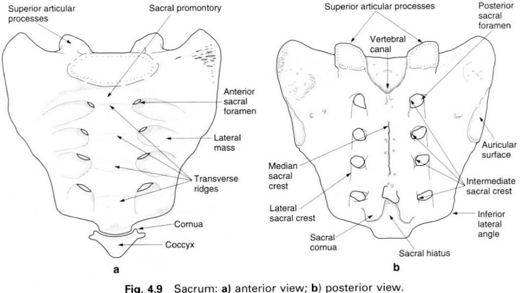
The dorsal surface of the sacrum is convex and
highly irregular, and presents the posterior
sacral foramina, medial to which the vertebral canal is closed over by the
fused laminae. However, usually the spinous processes and laminae of the fourth
and fifth sacral vertebrae are absent leaving the vertebral canal open. This is
the sacral hiatus, an inferior
entrance to the vertebral canal, which may be used, for example during labour,
to introduce an anaesthetics to block the sacral nerves. Posteriorally, in the
midline, the reduced spinous processes form the median sacral crest. Lateral to the dorsal sacral foramina are the
prominent lateral sacral crests,
representing the transverse processes. These lateral crests provide attachment
for the dorsal sacroiliac ligaments, and inferiorly for the sacrotuberous and
sacrospinous ligaments. Just medial to the dorsal sacral foramina are the
indistinct intermediate sacral crests,
representing the fused articular processes. The superior articular processes of the first sacral vertebra are large
and oval, being supported by short heavy pedicles. Their facets, for
articulation with the inferior articular surfaces of the fifth lumbar vertebra,
are concave from side to side, facing posteriorly and medially. The tubercles
of the inferior articular processes of the fifth sacral vertebral form the sacral cornua and are connected to the
cornua of the coccyx.
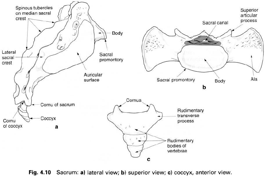
The lateral surface of the sacrum (figure a) is
triangular, being narrower below. The upper part of this surface is divided
into an anterior smoother pitted auricular
surface, covered in cartilage, for articulation with a similar area on the
ilium. The rougher posterior area presents three deep impressions for the
attachment of the powerful dorsal sacroiliac ligaments. The superior surface
(figure b) faces anterosuperiorly having a central oval area which is the upper
surface of the first sacral vertebra, being separated from the fifth lumbar
vertebra by a thick intervertebral disc. Its anterior projecting border is the sacral promontory. On each side of the
body of the sacrum is the ala, formed
by the fusion of the costal and transverse processes of the first sacral
vertebra. When articulated with the innominate, the ala of the sacrum is
continuous with the ala of the ilium.
Ossification
Primary centres appear in the sacrum between
the third and eighth month in utero;
one for each centre, one for each half of each vertebral arch, and for each
costal element in the upper four vertebrae. The costal elements fuse with the
arches by the age of 5 years, the arches with the centre slightly later, with
the two parts of each arch uniting between the ages of 7 and 10. The segments
of the lateral masses fuse together during puberty, with secondary centres
appearing for vertebral bodies at about the same time. Bodies and epiphyses
fuse between 18 and 25 years. Several secondary centres appear at the ends of
the costal and transverse processes from which two epiphyses are formed, one of
which covers the auricular surface while the other completes the lower margin
of the sacrum.


0 коментара:
Постави коментар