


The tarsal bones
The posterior talus
The
calcaneus
The calcaneus lies inferior to the talus and
projects backwards to form the prominence of the heel, and is strongly bound to
all the tarsal bones by ligaments. It is the largest bone in the foot, being oblong
in shape and having six surfaces. The anterior surface faces forwards for
articulation with the cuboid, being slightly convex from top to bottom and more
or less flat from side to side. The medial part of this surface extends on to
the medial side of the calcaneus to accommodate a backward projection of the
cuboid. The posterior surface is rounded, presenting three areas. The upper
part is smooth where a bursa lies between it and the tendocalcaneus. The
middle, which is smooth and convex, except at its lower margin where it ends as
a jagged rough edge, receives the attachment of the tendocalcaneus. The lowest
part is roughened, being subcutaneous and covered by the strong fibrous tissue
and fat of the heel pad. This lowest part of the posterior surface transmits
the body weight from the heel to the ground and curves forwards on to the
inferior surface.
Here are found the larger medial and smaller
lateral tubercles projecting forwards. The inferior surface continues forwards
as a rough area terminating as the anterior tubercle. The long plantar ligament
attaches to the rough area. The lateral surface of the bone is slightly
roughened and nearly flat. It presents two tubercles, one for the attachment of
part of the lateral ligament of the ankle joint, and the other, which is
slightly lower and more anterior, provides attachments for the inferior
peroneal retinaculum. The latter tubercle is elongated, with a groove above and
below, and is called the peroneal tubercle. The medial surface is smooth and
hollowed, being overhung anteriorly by the sustentaculum tali, under which is a
groove for the tendon of flexor hallucis longus. On the superior surface of the
sustentaculum tali is the middle articular surface for the head of the talus.
Behind the middle articular surface is a deep groove, the sulcus calcanei,
which continues across the superior surface of the calcaneus in a posteromedial
direction. In front of the sinus calcanei is a roughened area for the
attachment of muscles and ligaments, while behind it is the posterior articular
surface, convex from front to back and flat from side to side, for articulation
with the under surface of the body of the talus. Behind this articular surface
is a further roughened area, concave upwards from front to back and convex from
side to side.
The trabecular of the calcaneus have a
particular arrangement due to the weightbearing nature of the bone. From the
posterior articular surface, the supporting trabeculae pass downwards and
backwards to the heel and downwards and forwards to the articular area for the
cuboid. Running from the heel to the anterior surface are superior and inferior
arcuate systems serving to tie the bone together. Between all of these systems
there is an area of less dense, and therefore weaker, bone tissue.
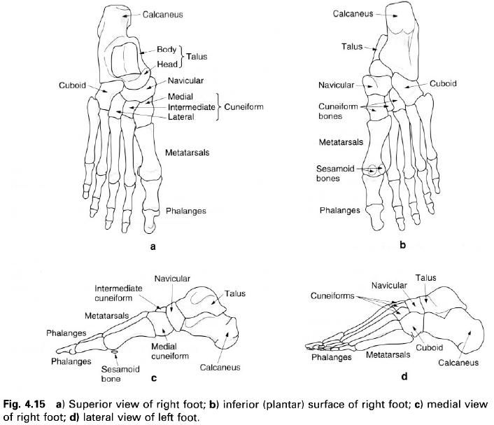
The
talus
The talus is situated above the calcaneus with
the head and neck directed forwards and medially. It transmits the body weight
from the tibia to the calcaneus and navicular. The body of the talus is
wedge-shaped from front to back being wider anteriorly, and lies between the
malleoli of the tibia and fibula. Its upper surface is convex from front to
back and slightly concave from side to side, being pulley-shaped, and
articulates with the trochlear surface of the tibia. The lateral surface of the
bone is triangular in shape with its apex pointing downwards, and articulates
with the medial surface of the lateral malleolus. The medial surface is partly
articular, with its upper articular part being comma-shaped. The medial and
lateral articular surfaces are continuous with the upper surface of the bone.
Below the medial articular surface is a depressed roughened area for attachment of the deep part of the
deltoid ligament. The inferior surface of the body is also articular, being
concave from front to back and articulates with the posterior facet on the
upper surface of the calcaneus.
At the posterior aspect of the bone, there is a
groove running downwards and medially for the tendon of flexor hallucis longus.
Lateral and medial to this groove are the lateral and medial tubercles of the
talus.
From the anterior and medial aspect of the body
the neck projects forwards and medially. Its upper, medial and lateral surfaces
are roughened, whilst its inferior surface presents an area for articulation
with the calcaneus on the upper surface of the sustentaculum tali. Just behind
the articular surface there is a deep groove termed the sulcus tali, which lies
immediately over the sulcus calcanei and forms with it the sinus tarsi.
The head of the talus is slightly flattened
anteriorly and articulates with the posterior surface of the navicular. Below
this main articulation there are two smaller articular areas, one for the upper
surface of the “spring” ligament and the other, which continues on to the inferior
surface of the neck, for the anterior articular area of the calcaneus.
The
navicular
The navicular lies anterior to the head of the
talus. On its inferomedial side it presents a large tuberosity. On its
inferomedial side it presents a large tuberosity. Its posterior surface is
concave for articulation with the head of the talus. Its anterior surface is
subdivided into three triangular areas by two faint ridges for articulation
with the three cuneiform bones. The inferior surface of the bone is narrow and
roughened for the attachment of ligaments and muscles. The small lateral and
subcutaneous upper surfaces are rough near their edges for the attachment of
interosseus ligaments, but together they form a curved surface.
The
cuboid
The cuboid has six surfaces, but in reality is
a cube that has been flattened from above downwards. It is situated on the
outside of the lateral cuneiform bone, in front of the calcanues and behind the
fourth and fifth metatarsals. Its posterior surface is slightly concave from
top to bottom, but flat from side to side, articulating with the anterior
surface of the calcaneus. Its medial surface is smooth on its anterior
two-thirds for articulation with the lateral cuneiform and sometimes the
navicular, whilst the posterior third is usually roughened for the attachment
of ligaments. Anteriorly, it is nearly flat, being divided by a slight ridge
into two facets for articulation with the bases of the fourth and fifth
metatarsals.
The lateral surface of the cuboid is by far the
smallest due to the convergence of the anterior and the posterior surfaces as
they pass laterally. Nearly the entire surface is taken up by a deep groove
passing downwards and forwards through which the tendon of the peroneus longus
passes. The groove is continued on the under surface of the bone and crosses
medially and anteriorly towards the medial cuneiform. The groove is very close
to the anterior border of the bone and is limited by a prominent ridge
posterior to it. The rest of the under surface of the cuboid is rough for the
attachment of the long and short plantar ligaments. Its dorsal surface is
roughened, and as in the case of the dorsal surfaces of the cuneiform bones and
navicular, is subcutaneous.
The
cuneiform bones
There are three cuneiform bones: the medial,
intermediate and lateral. As their name implies they are wedge-shaped, being
triangular at their anterior and posterior ends and having three rectangular
surfaces along their length.
The
medial cuneiform. The medial
cuneiform has its apex projecting upwards and its base downwards. Its anterior
and posterior surfaces are smooth for articulation with the first metatarsal
and the anterior surface of the navicular respectively. Its smooth lateral
surface articulates with the intermediate cuneiform in its posterior
two-thirds, and the base of the second metatarsal on its anterior third. Its
superior, medial and inferior surfaces form a continuous surface which forms
part of the medial side of the foot. This surface is roughened by ligaments and
has a smooth impression at the anteroinferior part of its medial aspect over
which the tendon of tibialis anterior runs. This is the largest of the three
cuneiforms.
The
intermediate cuneiform. The
intermediate cuneiform has its base uppermost and its apex projecting
downwards. It is shorter than the other two cuneiforms and is only
non-articular on its dorsal surface. It articulates medially with the medial
cuneiform, laterally with the lateral cuneiform, anteriorly with the second
metatarsal and posteriorly with the navicular. Part of the medial surface is
roughened for the attachment of ligaments.
The
lateral cuneiform. The apex of
the lateral cuneiform also projects downwards with its base uppermost. The
medial surface articulates mainly with the middle cuneiform having a small
facet anteriorly for articulation with the second metatarsal. Its lateral
surface articulates with the medial surface of the cuboid, its posterior
surface with the navicular and its anterior surface with the third metatarsal. The
nonarticular parts of the medial and lateral surfaces are roughened for the
attachment of ligaments.
The fact that the medial cuneiform has its base
projecting downwards, whilst the other two have their bases uppermost,
contributes to the arch shape across the foot from medial to lateral. With the
addition of the cuboid on their lateral side, the cuneiforms make up part of
the transverse tarsal arch.


0 коментара:
Постави коментар