


The irregularly shaped innominate consists of
three bones fused together: the ilium, the pubis and the ischium.
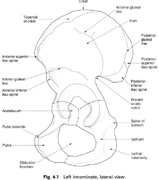
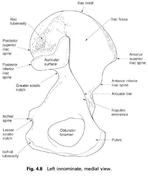
The ilium
The ilium is the upper broad blade for the
attachment of ligaments and large muscles. It forms the pelvic brim between the
hip joint and the articulation with the sacrum. The anterior two-thirds of the
projecting ilium forms the iliac fossa
medially, which is part of the lateral and posterior abdominal wall, and the
gluteal surface laterally, for attachment of the gluteal muscles. The posterior
one-third of the medial surface, which is thicker, carries the auricular surface for the articulation
with the sacrum; behind this is a prolonged rough part, the iliac tuberosity, for the attachment of
the strong sacroiliac ligaments which bear the body weight. The upper border of
the ilium is the iliac crest which is
convex superiorly as well as being curved anteroposteriorly with the anterior
part curved outwards. The iliac crest ends anteriorly as the anterior superior iliac spine and
posteriorly as the posterior superior
iliac spine. Both spines and the whole of the crest can be palpated. Behind
the anterior spine on the outer border is the prominent tubercle of the crest. Below the anterior superior spine is the anterior inferior iliac spine separated
by a shallow notch. Similarly, below the posterior superior spine is the posterior inferior iliac spine, again
separated by a shallow notch, the two posterior spines being closer together
than the two anterior spines. Below the posterior inferior spine is the deep greater sciatic notch. The iliac
fossa is separated from the sacropelvic surface of the ilium by the arcuate line which forms part of the
pelvic brim. The anterior part of this line has an elevation, the iliopubic eminence, marking the junction
of the ilium with the pubis. The part of the ilium which participates in the
formation of the acetabulum is the body of the ilium.
The outer gluteal surface of the ilium follows
the curvature of the iliac crest. It shows three curved gluteal lines which
demarcate the attachments of the gluteal muscles. The most obvious of these
lines is the posterior gluteal line,
which passes down from the iliac crest to the front of the posterior inferior
spine. The anterior gluteal line is a
series of low tubercles coming from the iliac crest curving upwards and
backwards below the iliac tubercle and then down towards the greater sciatic
notch. The inferior gluteal line is
less prominent, curving from below the anterior superior iliac spine towards
the apex of the greater sciatic notch. Below the inferior gluteal line is an
area of multiple vascular foramina. The fusion of the ilium and the ischium is
marked by a rounded elevation between the acetabulum and the greater sciatic
notch. Above this the ilium forms the major part of the notch. The gluteal
surface is succeeded inferiorly by the acetabular portion of the ilium.
The iliac fossa is the smooth internal
concavity of the ala of the ilium. It becomes narrower inferiorly ending at the
roughened iliopubic eminence, the line of junction between the ilium and the
pubis. Its deepest part, high in the fossa, is composed of paperthin
translucent bone. The pelvic brim, marked by the arcuate line of the ilium, is
the posterior – inferior limit of the iliac fossa. Behind and below the iliac
fossa and the arcuate line is the sacropelvic surface of the ilium. Posterior
to the iliac fossa, this region exhibits the auricular surface for articulation
with the first two segments of the sacrum and, behind and above it, the
tuberosity. The roughened tuberosity provides attachment for the short
posterior sacroiliac ligaments and for fibres of erector spinae and multifidus
muscles. The auricular area extends from the pelvic brim to the posterior
inferior iliac spine. Its surface is gently undulating, being convex above to
concave below, and roughened by numerous tubercles and depressions. The surface
is covered with hyaline cartilage forming a synovial joint, which is immobile,
with the ala of the sacrum. In later years, fibrous bands often joint the
articular surfaces within the joint space.
The pubis
The pubis is an angulated bone. The body of the
pubis projects laterally and superiorly as the superior ramus to join the ilium
and ischium at the acetabulum, forming one fifth of the acetabulum. A thin and
flattened inferior ramus extends inferiorly and posterolaterally from the body
to fuse with the ischium below the obturator
foramen. The body is quadrilateral in shape, with the symphyseal surface
being oval; it is crossed by several transverse ridges to which the
fibrocartilage of the symphysis pubis is attached, and coated with hyaline
cartilage for the secondary cartilaginous joint that constitutes the symphysis
pubis. The upper border of the body is the pubic crest, being marked laterally
by the pubic tubercle. From this
tubercle two ridges diverage laterally into the superior ramus. The upper of
these two ridges is the pectineal line, which is continuous with the arcuate
line of the ilium, and forms part of the pelvic brim. The lower, rounded ridge
is the obturator crest, passing downwards into the anterior margin of the
acetabular notch. Between these two ridges is the iliopubic eminence. Below the
obturator crest on the superior pubic ramus is the deep obturator groove.
The ischium
The ischium is the posterior inferior part of
the innominate, being angulated and in the same plane as the pubis. The apex of
the angulation is blunt and rounded, forming the ischial tuberosity, divided transversely by a low ridge. A smooth
oval above this ridge is further sub-divided by a vertical ridge into lateral
and medial areas. In the sitting positon, the weight of the body rests on the
two ischial tuberosities. Anteriorly the tuberosity passes upwards as the
ischial ramus, being continuous with the inferior pubic ramus, forming the
ischiopubic ramus. The body of the ischium forms two-fifths of the acetabulum.
The posterior border of the body is continuous above with the ilium, forming
the greater sciatic notch. Inferiorly this border ends as the blunt medially
projecting ischial spine, below which
is the groove forming the lesser sciatic
notch. The pelvic surface of the body is continuous with the pelvic surface
of the ilium, forming part of the lateral wall of the pelvis.
The acetabulum
is formed by the fusion of the three component bones of the innominate; the
ilium, ischium and pubis meet at a Y-shaped cartilage which forms their
epiphyseal junction. The anterior one-fifth of the acetabulum is formed by the
pubis, the superior posterior two-fifths by the body of the ilium, and the
inferior posterior two-fifths by the body of the ischium. The acetabulum is a
hemispherical hollow on the outer surface of the innominate, facing downwards,
forwards and laterally. The prominent rim of the acetabulum is deficient
inferiorly forming the acetabular notch. The rim gives attachment to the
acetabular labrum of the hip joint, its uneven internal edge provides an
attachment for the synovial membrane of the joint. The acetabular labrum
continues across the acetabular notch to produce the transverse ligament. The transverse ligament and margins of the
notch give attachment to the ligament of the head of the femur. The heavy wall
of the acetabulum consists of a semilunar articular portion covered with
hyaline cartilage, open below, and a deep, central non-articular portion, the
acetabular fossa. The acetabular fossa is formed mainly from the ischium, and
its wall is frequently thin.
The obturator foramen is a large aperture
ringed by the sharp margins of the pubis and ischium, those of the pubis
overlapping each other in a spiral forming the obturator groove, which runs
obliquely forwards and downwards from the pelvis into the thigh, being
converted into a canal by a specialization of the obturator fascia. The
obturator membrane is attached to the margins of the foramen, except superiorly
at the obturator groove.
Ossification
Each innominate bone ossifies from eight
centres: three primary centres, one each for the ilium, ischium and pubis; and
five secondary centres, one each for the iliac crest, the anterior inferior
iliac spine, the ischial tuberosity, the pubic symphysis and the triradiate
cartilage at the centre of the acetabulum. The sequence of ossification has
functional significance because of the support given to the pelvic organs and
its weight transmission role. The primary ossification centres appear during
the third, fourth and fifth months of development. At birth the ilium, ischium
and pubis are quite separate and the secondary ossification centres have not
yet appeared. By the age of thirtee or fourteen, the major parts of the ilium,
ischium and pubis are completely bony but are still separated by the Y-shaped
triradiate cartilage in the acetabulum. At the age of eight or nine, three
major centres of ossification appear in the acetabular cartilage. The largest
appears in the anterior wall of the acetabulum and fuses with the pubis.
Further centres appear in the iliac acetabular cartilage superiorly which fuses
with the ilium, and in the ischial acetabular cartilage posteriorly which fuses
with the ischium. Fusion of the three bones in the acetabulum occurs between
sixteen and eighteen years. The other secondary ossification centres appear at
about puberty and unite with the major bones between twenty and twenty-two.
Palpation
The anterior superior iliac spines can easily
be palpated in the living subject, particularly in females where they tend to
be further apart. They are situated at the anterior end of the iliac crest,
being found in the upper part of the pocket area. They act as an excellent
landmark for many of the surface markings in this region. Tracing backwards
from these spines, the iliac crest is easily palpable, having a large
tuberosity about 5cm from its anterior end. If this bony crest is followed as
far back as possible, the rather smaller posterior superior iliac spines can be
felt. Each is situated in a dimple in the female subject, whereas in the male
each spine appears as a small raised tubercle.
About 10cm below the centre of the iliac crest,
the greater trochanter of the femur can be clearly felt. Hip measurements, when
buying trousers or a skirt, are calculated erroneously around these two
trochanters. In sitting, the body rests on the ischial tuberosity of each
innominate bone; if the hands are
placed under this area the tuberosites can be easily felt. This part of the
tuberosity is covered with a bursa which often becomes painful and swollen when
sitting for too long on a hard surface; this is termed a bursitis.
If the hands are drawn down the front of the
abdominal wall, about 5cm above the genitalia, a bony ring can be felt. This
has a central depression where the pubic symphysis is situated with each pubic
tubercle about 1cm above and lateral on either side.


0 коментара:
Постави коментар