


The articulation is between the head of radius rotating within the fibro-osseous
ring formed by the radial notch of the ulna and the annular ligament.
Head of the radius
The beveled circumference of the head of the radius is covered by hyaline cartilage
continuous with that on its upper concave surface, so forming a smooth surface
for articulation with the ulna and
annular ligament(a). The anterior, medial and posterior parts of the
circumference tend to be wider than the lateral part, for direct articulation
with the ulna. The head of the radius tends not to be circular but is
slightly oval, with the major axis lying obliquely anteroposteriorly. The major
and minor axes have a length ratio of approximately 7:6.
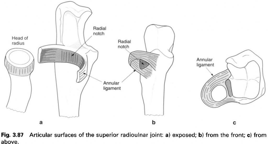
Radial
notch
The hyaline-covered radial notch is continuous
with the trochlear notch of the ulna
on its lateral side, being separated from it by a blunt ridge(a,c). It forms
approximately one-fifth of the articular fibro-osseous ring, and is therefore
concave anteroposteriorly but almost flat vertically.
Annular
ligament
The flexible annular ligament forms the
remaining four-fifths of the articular surface which encircles the head and
neck of the radius. Its flexibility
enables the oval head of the radius
to rotate freely in pronation and supination. The ligament is a strong,
well-defined band attached to the anterior and posterior margins of the radial
notch of the ulna. Posteriorly the
ligament widens where it attaches to adjacent areas of the ulna above and below the posterior margin of the notch. The
diameter between its lower borders is narrower than that above(c), so it cups
in under the head of the radius and
acts as a restraining ligament preventing downward displacement of the head
through the ring.
Superiorly the annular ligament is supported by
the firm fusion of the radial collateral ligament and the blending of the
lateral part of the fibrous capsule of the elbow joint in front and behind.
Inferiorly a few loose fibres attach the ligament to the neck of the radius beyond the epiphyseal line. These
fibres are too loose to interfere with movements at the joint, but give some
support to a dependent fold of synovial membrane. The upper part of the
ligament is lined with fibrocartilage continuous with the hyaline cartilage of
the radial notch. The lower part of the ligament is lined with synovial
membrane.
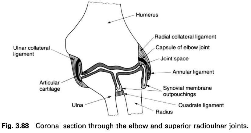
Joint capsule and synovial membrane
The superior radioulnar joint is continuous
with the elbow joint and consequently shares the same joint capsule. The
synovial membrane associated with the elbow part of the joint space attaches to
the upper margin of the fibrocartilage lining of the annular ligament. From the
lower border of the fibrocartilage, and lining the lower part of the annular
ligament, the synovial membrane extends below the lower border of the ligament
to hang as redundant fold which has a loose attachment to the neck of radius. The membrane lies on the upper
surface of the quadrate ligament, which limits and supports it, and passes
medially from the radius to attach to
the lower border of the radial notch of the ulna.
The redundancy of the synovial membrane below the annular ligament accommodates
to the twisting of the membrane that accompanies rotation of the radius.
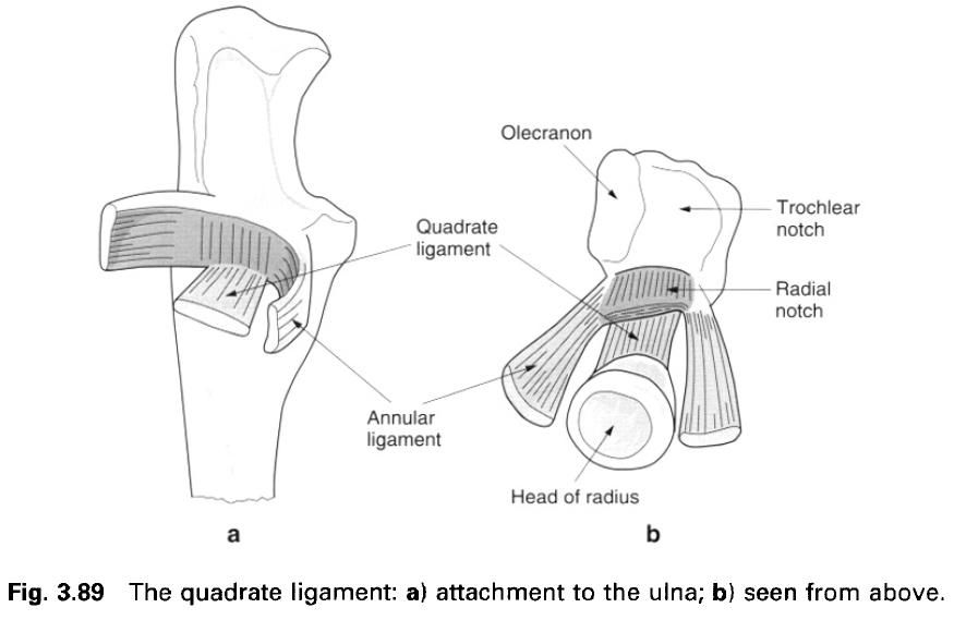
Ligaments
Although the annular ligament provides an
important support for the head of the radius,
it is not sufficient by itself to provide the only support to the superior
radioulnar joint, because of its need to change shape with rotation of the radius. Indeed, this constant need to
accommodate to the changing orientation of the head of the radius may lead to stretching of the ligament. Consequently, there
are additional structures which provide support to the joint: the quadrate
ligament and the interosseus membrane.
Quadrate
ligament
The quadrate ligament stretches from the lower
border of the radial notch of the ulna
to the adjacent medial surface of the neck of the radius proximal to the radial tuberosity. Its fibres run in a
criss-cross manner between the two bones, so that, irrespective of the relation
of the radius to the ulna, some fibres are always under
tension. The overall tension within the ligament thus remains constant in all
positions of pronation and supination. Its two borders are strengthened by
fibres from the lower border of the annular ligament.
Blood and nerve supply
The arterial blood supply to the superior
radioulnar joint is by branches from vessels supplying the lateral part of the
elbow joint; namely the middle and radial collateral branches of the profunda
brachii, and the radial and interosseus recurrent branches from the radial and
common interosseus arteries respectively. Venous drainage is by similarly named
vessels draining eventually to the brachial vein. Lymphatic drainage is by
vessels traveling with the arteries to small nodes associated with the main
arteries and then to the lateral group of axillary nodes.
The nerve supply to the joint is by twigs from
the posterior interosseus branch of the radial nerve, the musculocutaneous and
median nerves, with a root value of C5, 6 and 7.
Surface
marking and palpation
The line of the superior radioulnar joint can
be palpated posteriorly. Having identified the head of the radius in the depression on the posterolateral aspect of the elbow,
a vertical groove between the radius
and ulna can be felt medially. This
is the position of the joint line. During pronation and supination, the head of
the radius can be felt rotating
against the ulna.
Relations
Anteriorly the joint is crossed by the tendon
of biceps passing to its attachment
of the radial tuberosity; posteriorly is the fleshy belly of anconeus . Medial to the tendon of biceps lies the brachial and then the
radial artery from above downwards.
Stability
The joint has a reasonable degree of inherent
stability. However, in children, the head of the radius may be pulled from the confines of the annular ligament in
traction dislocation. Tears of the annular ligament will also result in
dislocation at the joint.
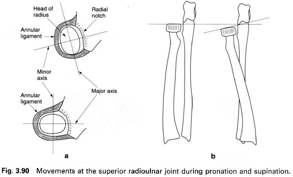
Movement
The main movement that occurs at the superior
radioulnar joint is rotation of the head of the radius within the fibro-osseous ring of the annular ligament and
radial notch of the ulna(a). The
movement is probably limited by tension developed in the quadrate ligament.
In addition to this principal movement, there
are four other movements related to the joint. These are:
1)
Rotation
of the superior concave surface of the radial head in relation to the capitulum
of the humerus;
2)
The
beveled ridge of the radial head glides in contact with the capitulotrochlear
groove of the humerus;
3)
The head
of the radius is displaced laterally
because the major axis of the oval head comes to lie transversely(a);
4)
The plane
of the radial head becomes tilted laterally and inferiorly during pronation due
to the radius moving obliquely around
the ulna(b).
Accessory
movements
Gripping the head of the radius between the thumb and index finger, it can be moved
anteroposteriorly with respect to both the ulna
and the capitulum.


0 коментара:
Постави коментар