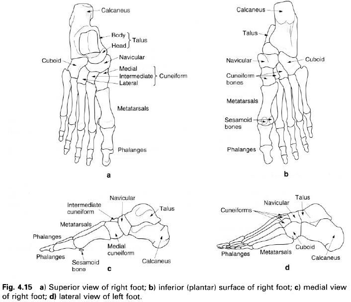
Free Facebook Likes, Youtube Subscribers,  Twitter Followers

Ads 468x60px

Blogger Tricks

Blogger Themes

29. 3. 2013.

Evaluating the center of gravity of dislocations in soccer players with and without reconstruction of the anterior cruciate ligament using a balance platform

OBJECTIVE: The objective of this study was to compare the dislocation of the center of gravity and postural balance in sedentary and recreational soccer players with and withoutanterior cruciate ligament (ACL) reconstruction using the Biodex Balance System (BBS).
METHOD: Sixty-four subjects were divided into three groups: a) soccer players who were post- anterior cruciate ligament reconstruction; b) soccer players with no anterior cruciate ligament injuries; and c) sedentary subjects. The subjects were submitted to functional stability tests using the Biodex Balance System. The instability protocols used were level eight (more stable) and level two (less stable). Three stability indexes were calculated: the anteroposterior stability index, the mediolateral stability index, and the general
stability index for all the subjects of the experiment.
RESULTS: Postural balance (dislocation) on the reconstructed side of the athletes was worse than on the side that had not undergone reconstruction. The postural balance of the sedentary group was dislocated less on both sides than the reconstructed knees of the athletes without anterior cruciate ligament injuries. There were no differences in postural balance with relation to left/right dominance for the uninjured athletes and the sedentary individuals.
CONCLUSION: The dislocation of the center of gravity and change in postural balance in sedentary individuals and on the operated limb of Surgery Group are less marked than in the soccer players from the Non Surgery Group and on the non-operated limbs.
The dislocation of the center of gravity and the change in postural balance from the operated limb of the soccer players is less marked than in their non-operated limbs.


28. 3. 2013.

Proprioceptive neuromuscular facilitation training induced alterations in muscle fiber type and cross sectional area


Objectives: To compare the effects of proprioceptive neuromuscular facilitation(PNF) and isokinetic training on fibre type distribution and cross sectional area of the vastus lateralis muscle.
Methods: Twenty four university students, males, were divided into two groups: PNF training and isokinetic training(ISO). The training regimen for the PNF group consisted of 3 sets of 30 repetitions against maximal resistance, alternating two patterns of sequential movements of the right lower extremity: a) toe flexion and ankle plantar flexion and eversion; b) knee extension and hip extension, abduction and internal rotation. The ISO group performed three sets of 30 repetitions alternating knee extension and flexion of the right leg at angular velocities of 180 and 90° /s in an isokinetic dinamometer (Cybex). Both groups trained three times a week for a total time of 8 weeks. Muscle biopsy specimens were obtained from the right vastus lateralis muscle before and after training.
Results: The mean percentage area of the type IIB fibre was significantly decreased (p<0.01) after eight weeks of PNF training, whereas that of type IIA fibre was significantly (p<0.05) increased. The mean percentage area of ISO trained type IIAB fibres exhibited in augmentative pattern (p<0.01) with a parallel reduction (p<0.05) in type IIA. Percentage fibre type distribution exhibited a similar pattern.
Conclusions: Both PNF and ISO training alter fibre type distribution and mean cross sectional area. These changes occur in type II fibre subgroup.   

26. 3. 2013.

Muscles abducting the hip joint


Gluteus medius
Gluteus minimus
Tensor fascia latae

Gluteus medius

Gluteus medius is situated on the lateral and upper part of the buttock, just below the iliac crest. It must be considered to be a close companion of gluteus maximus and is in fact overlapped by this muscle from the back. It is fan-shaped, having its broad part above and its tendon below. It fills the space between the iliac crest and the greater trochanter of the femur.
Its upper attachment is to the gluteal, or lateral surface of the ilium between the posterior and anterior gluteal lines. This area is quite extensive reaching to the iliac crest above and almost as far as the sciatic notch below. The muscle is covered with a strong layer of fascia from the deep surface to which it has a firm attachment. It shares the posterior part of this fascia with gluteus maximus.
The posterior fibres pass downwards and forwards, the middle fibres pass straight downwards and the anterior fibres pass downwards and backwards. The fibres come together and form a flattened tendon which attaches to a roughened area, which runs downwards and forwards, visible on the superolateral side of the greater trochanter of the femur. The tendon is separated from the trochanter by a bursa, whose position is given by a smooth area on the trochanter in front of the attachment of the tendon.

Nerve supply

Gluteus medius is supplied by the superior gluteal nerve, root value L4, 5, S1. The skin covering the muscle is mainly supplied from L1, 2.

Action

With the pelvis fixed, gluteus medius will pull the greater trochanter of the femur upwards. However, as the fulcrum of the movement is at the hip joint, this will cause the femoral shaft to move laterally, this is termed abduction.
If the lower attachment of the muscle is fixed it will pull down the wing of the ilium, producing a downward tilting of the pelvis to the same side and, of course, a raising of the pelvis on the opposite side. In additioin, the anterior fibres of gluteus medius acting from a fixed pelvis will help with medial rotation of the femur. Acting with the femur  fixed, these fibres rotate the opposite side of the pelvis forward.

Functional activity

Gluteus medius plays a vital role in walking, running and when bearing weight on one limb. When the opposite limb is taken off the ground the pelvis on that side would tend to drop through loss of support from below. Gluteus medius on the supporting side works very hard to maintain, or even raise a little, the opposite side of the pelvis, allowing the raised limb to be brought forward for the next step. If the muscle is paralysed the pelvis drops on the opposite side during this manoeuvre.
In walking or running, not only is gluteus medius important for support, but with the help of other muscles, such as gluteus minimus and tensor fascia lata, it produces a rotation of the hip joint. This time with the femur the more fixed point, it controls the pelvic rotation on the same side.
If the muscle is unable to work efficiently due to paralysis or poor mechanics of the hip joint, the pelvis will drop on the opposite side. This is reffered to as a Trendelburg sign. Walking in the case is awkward and difficult, and running virtually impossible.

Palpation

Find the middle of the iliac crest, which is directly above the greater trochanter of the femur. About two fingers’ breadth below this region is the bulk of the muscle. Now stand alternately on one limb and then the other; you will feel the muscle become hard as the weight is borne on the same limb. Place the fingers of the other hand on the opposite side; walk slowly down the room. You will feel the two muscles coming into action alternately.
A patient with a Trendelburg gait, either on one or both sides, compensates for the lack of support of the swing limb by throwing the trunk over the supporting limb so that the weight is balanced over the hip, thus giving time to swing the limb through.


Gluteus minimus

Although this is the smallest of the gluteal muscles it takes the largest attachment from the gluteal surface of the ilium. It is triangular in shape, being wide at the top and narrowing to a tendon below.
Its upper attachment is from the gluteal surface of the ilium in front of the anterior and above the inferior gluteal lines, reaching as far forward as the anterior border of the ilium in front and almost to the sciatic notch behind. Its fibres pass downwards, backwards and slightly laterally forming a tendon which attaches to a small depression on the anterosuperior aspect of the greater trochanter of the femur.

Nerve supply

Gluteus minimus is supplied by the superior gluteal nerve, root value L4, 5, S1. The skin overlying the muscle is mainly supplied by L1.

Action

If the upper attachment of the muscle is fixed, contraction of its anterior fibres will medially rotate the femur. This is because the femoral attachment lies lateral to the fulcrum of the movement, the hip joint. If the lower attachment is fixed, the muscle will raise the opposite side of the pelvis in a similar way to gluteus medius. It will also, by pulling the front of the ilium outwards, swing the opposite side of the pelvis forwards.

Functional activity

This muscle appears to play its most important role in the support and control of pelvic movements. It is a well-developed and powerful muscle, using its power to a maximum in walking and running when the opposite limb is off the ground. As the limb is swung forward, the pelvis on the same side is also swung forward. This uses the hip of the weight-bearing limb as the fulcrum of the movement, with gluteus medius and minimus both supporting the pelvis and swinging it forward on the opposite side.

Palpation

Find the anterior superior iliac spine at the front of the iliac crest. Allow the pads of your finger to slip downwards and backwards towards the greater trochanter of the femur. Within two fingers’ breadth you will be on the muscle bulk. Now rotate the lower limb medially and you will feel the muscle belly contracting hard. Do the same on the opposite side of the body and then begin to walk forward. You will feel the muscles contracting alternately, as each limb becomes weight-bearing.

Tensor fascia latae

Tensor fascia latae is situated anterolateral to the hip joint and superficial to gluteus minimus. It attaches above to the anterior part of the outer lip of the iliac crest, between and including the iliac tubercle and the anterior superior iliac spine, the area of gluteal surface just below it, the fascia between it and gluteus minimus and that covering its superficial surface. Inferiorly, it attaches between the two layers of the iliotibial tract, below the level of the greater trochanter. 


Nerve supply

Tensor fascia latae is supplied by the superior gluteal nerve, root value L4, 5, with the skin overlying the muscle supplied by L1.

Action

This muscle overlies gluteus minimus and helps in flexion, abduction and medial rotation of the hip joint. It also straightens out the backward pull of gluteus maximus on the iliotibial tract.
Acting with the superficial fibres of gluteus maximus it will tighten the iliotibial tract, and through its attachment to the lateral condyle of the tibia, will extend the knee joint. Acting with gluteus minimus it will medially rotate the hip joint and its posterior fibres may help in abduction of the thigh.

Functional activity

Due to the fact that this muscle, together with gluteus maximus, links the pelvis with the tibia it will help to steady and control the movements of the pelvis and femur on the tibia when the limb is weight-bearing. Tensor fascia latae produces strong medial rotation when the hip is in extension and the lower limb, pelvis and trunk are prepared to take the thrust relayed through the lower limb by the calf muscles during the “toe-off” phase of walking.
When the quadriceps femoris is paralysed, tensor fascia lata can be developed to produce sufficient extension of the knee to enable the patient to walk, but its action is only weak and limited in range.

Palpation

Place the fingers half way between the anterior superior iliac spine and the greater trochanter of the femur. When the lower limb is medially rotated, the muscle can be felt to contract powerfully. If the weight is taken on the limb and the pelvis is rotated to the same side, a similar contraction of the muscle will be observed.

25. 3. 2013.

Muscles extending the hip joint


Gluteus maximus
Semitendinosus

HAMSTRINGS
Semimbranosus
Biceps femoris

Gluteus maximus

As its name implies, this is the largest of the gluteal muscles. It is very powerful and is situated on the posterior aspect of the hip joint, being responsible for the very pleasant shape of this region in humans. In lower primates gluteus maximus in an adductor of the hip; this was also the case in early, primitive humans. However, with the changes that have occurred in the human pelvis to enable the erect posture to be adopted, the muscle has become mainly an extensor of the hip. It is the muscle mainly responsible for the erect position, thereby freeing the forelimbs from a weight-bearing role, and enabling them to become the precision implements that they are today.
Gluteus maximus is quadrilateral in shape consisting of bundles of muscle fibers laid down in the line of pull of the muscle, giving the surface of the muscle a coarse appearance. The muscle is thick and forms two layers as it passes down to its lower attachment. Above, it attaches to the gluteal surface of the ilium behind the posterior gluteal line, the posterior border of the ilium and from the adjacent part of the iliac crest. It also arises from the side of the coccyx and the posterior aspect of the sacrum, including the upper part of the sacrotuberous ligament. Its upper fibres attach to the aponeurosis of the sacrospinalis while its deep anterior fibres come from the fascia which covers gluteus medius. Although this appears to be a vast area, it must be remembered that most of these structures can be covered with one hand if it were slipped into the back pocket of a pair of trousers.
The fibres pass downwards and forwards towards the upper end of the femur. The most superficial, about three-quarters, of the fibres form a separate lamina, which narrows down and attaches between the two layers of the fascia lata helping to form the iliotibial tract. The deeper remaining one-quarter of the muscle fibres form a broad aponeurosis which attaches to the gluteal tuberosity of the femur


Nerve supply

Gluteus maximus is supplied by the inferior gluteal nerve, root value L5, S1, 2. The skin covering the muscle, however, is mainly supplied by branches from L2 and S3.

Action

When acting from above, the muscle pulls the shaft of the femur backwards producing extension of the flexed hip joint. As its lower attachment is nearer to the lateral side of the thigh, the muscle will tend to rotate the thigh laterally during extension. The lower fibres can adduct the thigh, while the upper fibres may help in abduction.
The fibres which attach to the iliotibial tract can produce extension of the knee joint because the lower end of the tract attaches to the lateral tibial condyle anterior to the axis of movement. Through the iliotibial tract, gluteus maximus provides powerful support on the lateral side of the knee. If the femur is fixed, contraction of gluteus maximus will pull the ilium and pelvis backwards around the hip joint, but this time the pelvis and trunk are the moving parts, and a lifting of the trunk from a flexed position occurs.

Functional activity

Being a powerful extensor of the thigh, especially when the hip has been flexed, means that this muscle is ideally suited for fulfilling its role in such powerful movements as stepping up onto a stool, climbing and running. However, it is not used greatly as an extensor in ordinary walking.
With the hamstrings, it will take part in raising the trunk from a flexed position, as in standing upright from a bent forward position. Indeed, gluteus maximus and the hamstrings provide the main control in forward bending of the body, as the movement primarily occurs at the hip joint.
It plays an important role in balancing the pelvis on the femoral heads thus helping to maintain the upright posture; its ability to aid lateral rotation of the femur when standing assists in raising the medial longitudinal arch of the foot.
The role of gluteus maximus during sitting should not be dismissed. Although the ischial tuberosities support the majority of the weight of the trunk when sitting, pressure is regularly relieved from these bony points by a static or sometimes dynamic contraction of the muscle which raises the tuberosities of the ischium from the supporting surfaces. The muscle is then relaxed and the weight is then lowered. Sometimes the weight is shifted from side to side with the alternate use of gluteus maximus of each side.
Paralysis of the muscle will lead to a flattening of the buttock with a loss of the beautiful contour, and an inability to climb stairs and run. However, it must be kept in mind that there are other muscles which can be brought into action to produce extension of the hip, although it is much weaker movement. Gluteus maximus can be developed to produce a functional extension of the knee in patients where quadriceps femoris is either very weak or paralysed. This is not a powerful movement, but may be sufficient to enable the patient to extend the knee and enable the lower limb to become weight-bearing during walking or standing.

Palpation

On a model or on yourself, first find some bony prominences which will give useful landmarks of the muscle. The iliac crest is easily palpated approximately at the belt level; moving the hand backwards along the crest a small bony process can be felt: this is the posterior superior iliac spine. With the fingers running inferiorly and medially let this be the centre of the palm. The hand will now just about cover the upper attachment of gluteus maximus; the palm is over the posterior part of the ilium, the sacrum and the back of the sacroiliac joint, while the tips of the fingers are on the edge of the coccyx and the upper end of the sacrotuberous ligament. The bulk of the muscle is now under the palm, follow this path to the greater trochanter of the femur. Now try the following:
  1. Extend the lower limb whilst in the standing position, keeping the hand on the muscle; it goes hard and produces a much clearer shape.
  2. Place the foot onto a stool and put the hand in the same position as before and step up. Again the muscle will be felt coming into action very strongly.
  3. Take up the standing position and place the hands on each gluteus maximus as if they were in the back pocket. Raise the medial borders of the feet as if to shorten the medial longitudinal arch of the foot. As the arch is raised gluteus maximus will be felt working quite strongly, with the femur tending to rotate laterally.
  4. Finally, take up the sitting position but this time place a hand under each buttock so that the ischial tuberosity now rests on the hand. Now move the weight from side to side as if getting tired of sitting. Gluteus maximus now contacts alternately, taking the weight off the tuberosity and then lowering it down again.

Semitendinosus

The upper attachment of this muscle is from the lower medial facet of the lateral section of the ischial tuberosity. Its tendon of attachment is combined with that of the biceps femoris and the two muscles run together for a short distance. It then forms a fusiform muscle belly which quickly gives way to a long tendon, thus accounting for its name. This tendon passes downwards and medially behind the medial condyle of the femur, being separated from the medial collateral ligament by a small bursa, to attach to a vertical line on the medial surface of the medial condyle of the tibia just behind the insertion of the sartorius and behind and below the attachment of the gracilis. Near its insertion it is separated from gracilis is separated from sartorius by another bursa. 


Nerve supply. Semitendinosus is supplied by the tibial division of the sciatic nerve, root value L5, S1, 2. The skin covering the muscle is supplied mainly by S2.
Action. Semitendinosus will, when working from below, help to extend the hip joint when the trunk is bent forward. When working from above it will aid in flexion of the knee joint; if the knee is semiflexed it will produce medial rotation of the knee. If the foot is fixed, semitendinosus will act as a lateral rotator of the femur and pelvis on the tibia.

Semimembranosus

This muscle is situated on the posteromedial side of the thigh in its lower part, deep to semitendinosus. It attaches by a strong membranous tendon to the upper lateral facet on the rough part of the ischial tuberosity and passes downwards and medially. The muscle becomes fleshy on the medial side of the tendon, being deep to semitendinosus and biceps femoris. From the lower part of the muscle a second aponeurotic tendon arises narrowing down towards its lower attachment, which is a horizontal groove on the posteromedial surface of the medial tibial condyle. From here its fibres spread in all directions, but particularly upwards and laterally forming the oblique popliteal ligament. Bursae separate the muscle from the medial head of gastrocnemius and from the tibia near its attachment.


Nerve supply. Semimembranosus is supplied from the tibial division of the sciatic nerve, root value L5, S1, 2. The nerve supply to the skin covering the muscle is the same as that for the semitendinosus, that is mainly from S2.
Action. As for semitendinosus.

Biceps femoris

 Biceps femoris is situated on the posterolateral aspect of the thigh, arising by two heads as the name implies, these being separated by a considerable distance.
The long head attaches to the lower medial facet on the ischial tuberosity with the tendon of the semitendinosus, spreading on to the sacrotuberous ligament. These two tendons descend together for a short distance then separate into the two individual muscles, the long head of biceps forming a fusiform muscle running downwards and laterally across the posterior aspect of the thigh superficial to the sciatic nerve. In the lower third of the thigh the long head begins to narrow and is joined on its deep aspect by the short head of biceps.
The short head has its upper attachment from the lower half of the lateral lip of the linea aspera reaching almost as far up as the attachment of gluteus maximus and running down onto the upper half of the lateral supracondylar line of the femur; some fibres arise from the lateral intermuscular septum. The fibres of this short head gradually blend with the narrowing tendon of the long head which lies superficial to it.
On approaching the knee, the tendon can be felt crossing its posterolateral aspect running towards the head of the fibula


Prior to its attachment to the head of the fibula the tendon of biceps femoris is split in two by the fibular collateral ligament. Some fibres of the tendon join the ligament, while a few others attach to the lateral tibial condyle and some to the posterior aspect of the lateral intermuscular septum which lies just in front of it. A bursa separates the tendon from the fibular collateral ligament.
Nerve supply. The long head of biceps femoris is supplied by the tibial portion of the sciatic nerve, while the short head is supplied by the common peroneal portion; the root value of both is L5, S1, 2. The skin covering the muscle is supplied mainly by S2.
Action. Biceps femoris helps the previous three muscles to extend the hip joint, particularly when the trunk is bent forwards and is to be raised to the erect position. All three hamstrings will, of course, control the lowering forward of the trunk; however in this case they are working eccentrically, that is working, but its two ends moving apart. Biceps femoris aids the semimembranosus and semitendinosus muscles in flexing the knee joint. With the knee in a semiflexed position biceps femoris will rotate the leg laterally on the thigh or if the foot is fixed, medially rotate the thigh and pelvis on the leg.

The hamstrings

Semitendinosus, semimbranosus and biceps femoris are collectively known as the hamstrings.
They make up the large mass of muscle which can be palpated on the posterior aspect of the thigh, and are involved with extension of the hip, flexion of the knee and rotation of the flexed knee. When working from above they can either flex the knee as a group, or they can work individually in rotating the flexed knee. If they are working with the lower attachment fixed, they will act as a group extending the hip joint.

Functional activity of the hamstrings

All three muscles cross the posterior aspect of both the hip and the knee joints. Flexion of the knee and their stabilizing effect is no doubt a very important function of these muscles, although for this action a much smaller muscle bulk would have been sufficient. Extension of the hip joint when the thigh is the moving part would also require a far smaller group of muscles, especially when it is remembered that gluteus maximus is far better situated to do the job. Raising the trunk from a flexed position on the other hand requires a great deal more power as the muscles are working with a very short lever arm: the ischium and its ramus. The weight of the trunk acting on the other side of the hip joint is considerable.
The mode of action of this group of muscles may well be the reason it is injured so frequently during sports activities. The most common cause of sports injury appears to be in the running section of athletics, being more common in the first 10-20m of a sprint. This is often blamed on inadequate preparation and warm-up before the start, and to some extent this may be true. It must, however, be remembered that at this stage in a race the hamstrings are contracting strongly and are acting over two joints.
At the start of a race the athlete is in a forward-lean position in order to gain as much forward motion as possible. Starting blocks serve to increase the degree of forward leaning. The hamstrings are therefore working to their maximum, either to raise the trunk to an upright position, or to hold the trunk in such a position that forward collapse of the body as a whole is imminent. At the same time the lower limb is being thrust forward to gain as much ground as possible, with flexion of the knee to prevent the foot touching the ground. The hamstrings must be under immense strain in this position and it is not surprising that the muscle may tear.
The hamstrings play an important part in the fine balancing of the pelvis in the standing position, particularly when the upper trunk is being moved off the vertical axis. Working in conjunction with the abdominal muscles anterosuperiorly and gluteus maximus posteroinferiorly, the anteroposterior tilt of the pelvis can be altered. This will have an effect on the lumbar lordosis.
Finally, the hamstrings  have a role in decelerating the forward motion of the tibia when the free swinging leg is extended during walking, and so prevents the knee snapping into extension.

Muscles around the hip joint


Deep within the gluteal region is situated the hip joint. This is a ball and socket joint capable of movement in many directions.
To produce these movements there is a complex arrangement of muscles around the joint which either act on the thigh with respect to the thigh. It must be remembered that during many of these movements the hip joint is weight-bearing, transmitting the weight of the body above it, via the lower limbs, to the ground.
Thus, the muscles surrounding the joint have a dual role. They must be capable of immediate controlled power when needed for sudden powerful activities such as running uphill or upstairs, and yet retain the ability to maintain a set position for long periods of time as in standing, leaning forwards and even sitting.
The hip joint is surrounded on all sides by muscles, which are much thicker and stronger around the posterior and lateral aspects and consequently the joint appears to be situated near the front of the region.
Muscles anterior to the joint tend to be flexors, those posterior tend to be extensors, medially they tend to be adductors and laterally abductors. Both medial and lateral rotation occurs at this joint because of the obliquity of some of the muscle fibres. This is explained more fully under the individual muscles.
Some of the muscles in this region will have their effect on more than one joint. When this is the case, reference is made to the muscle in both regions, but details are only found in one section.

19. 3. 2013.

The bony structure of the foot - part III


THE METATARSALS

There are five metatarsal bones in each foot, the most medial of which is by far the stoutest, although it is also the shortest. The second metatarsal is the longest, whilst the fifth can be recognized by the large tubercle which projects backwards and laterally from its base. All five metatarsals have certain features in common: a shaft, with a head distally, and a base at the proximal end. The bases articulate with the tarsal bones while the heads articulate with the proximal phalanx of each toe.
The base of the first metatarsal is concave from side to side and flat from top to bottom, articulating with the anterior surface of the medial cuneiform. Its lateral surface has a facet for articulation with the base of the second metatarsal, whilst its inferior surface projects downwards ending as a tuberosity. The base of the second metatarsal articulates with the intermediate cuneiform posteriorly. Medially it articulates with the medial cuneiform and the first metatarsal, and laterally with the lateral cuneiform and the third metatarsal. The base of the third metatarsal is flat and articulates with the lateral cuneiform, and on either side with the adjacent metatarsals. It is roughened on its upper and lower surfaces. The fourth and fifth metatarsal bases articulate with the anterior surface of the cuboid. The fourth has a small facet on either side for articulation with the adjacent metatarsals, whereas the fifth base is more expanded, having a large tubercle on its lateral side. The upper and lower surfaces of each are roughened.
All the shafts are more or less cylindrical in form, the first being the thickest and the second usually being the thinnest. All, however, become narrower as they pass forward towards their heads.
The heads are smooth, convex from above downwards as well as from side to side. On either side, just behind the head, is a tubercle in front of which is a small depression for the attachment of ligaments. The superior non-articular surface is roughened, while the inferior surface is marked by a groove passing forwards, which gives passage to the long and short flexor tendons. The head of the first metatarsal is large and wide forming the ball of the great toe. It articulates with the base of the first phalanx and two sesamoid bones. The plantar surface of this bone is grooved, on each side of a prominent central ridge, by the sesamoid bones in the tendons of the short muscles which pass inferior to it.


THE PHALANGES

There are two phalanges in the great toe and three in each of the other toes. They are miniature long bones having a shaft and two extremities and with certain features in common. Each of the bases of the proximal phalanges has a proximal surface which is smooth and concave for articulation with the head of its metatarsal. The remaining phalanges have a proximal surface divided into two by a vertical ridge. Each bone is flattened on its plantar surface and rounded on its dorsum. The head of each bone, except the terminal phalanges, is divided into two condyles by a vertical groove giving it a pulley shape. The articular surface tends to be more extensive on the plantar surface of the head where it joins the flattened surface of the shaft. The sides of the heads are roughened, being marked by a small tubercle at the centre.
The head of each distal phalanx is flattened on its dorsum and has no articular area. This surface is, of course, the nail-bed.

Ossification of the bones of the foot

Each tarsal bone ossifies from a primary centre which appears in the cartilaginous precursor. The calcaneus is the only tarsal bone to have a secondary centre. The primary centres for the calcaneus and talus appear before birth in the sixth and eighth months in utero respectively. That for the cuboid appears at nine months in utero and may therefore be present at birth; if not, it appears soon afterwards. The centres of ossification for the remaining bones appear as follows: at the end of the first year for the lateral cuneiform and navicular, and during the fourth year for the intermediate cuneiform. Ossification of these bones is completed shortly after puberty.
The secondary centre for the calcaneus appears at about 9 years in its posterior end and extends to include the medial and lateral processes. Fusion occurs between 15 and 20 years. Occassionally, the lateral tubercle may ossify separately.
Because the ossification centres for the calcaneus, talus and cuboid are usually present before birth, they can be used to assess the skeletal maturity of a new-born child. They may be used in conjunction with the secondary centres in the distal end of the femur and in the proximal end of the tibia.

The metatarsals

A primary centre appears in the body of each metatarsal at 9 weeks in utero, so that at birth they are well ossified. Secondary centres appear in the base of the first metatarsal and in the heads of the remaining metatarsals during the second and third years, with the medial ones appearing earlier. Fusion of the epiphyses with the bodies occurs between 15 and 18 years. In the lateral metatarsals, the epiphyses may occasionally be found in the bases instead of  the heads.

The phalanges

The primary centres for the distal and proximal phalanges appear during the fourth month in utero, with the distal ones appearing first. The primary centre for the middle phalanx appears between 6 months and birth. Secondary centres for the bases of all phalanges appear during the second and third years and fuse with the bodies between 15 and 20 years.
It is interesting to note that the first metatarsal has an ossification pattern similar to that of the phalanges. It could be argued that instead of the middle phalanx being missing in the great toe it is the metatarsal, so that what we now refer to as the first metatarsal is in fact an enlarged proximal phalanx.

Palpation of the bones of the foot

Posteriorly, the calcaneus can clearly be identified being subcutaneous on its lateral, posterior and medial aspects. The inferior surface is covered with thick plantar fascia, but the medial and lateral tubercles are identifiable on deep palpation posteriorly. Medially, 1cm below the tip of the medial malleolus, the sustentaculum tali appears as a horizontal ridge,  whilst on the lateral aspect, the peroneal tubercle lies approximately 2cm below and in front of the tip of the lateral malleolus, with the lateral tubercle of the calcaneus( for the attachment of the calcaneofibular ligament) a little behind and above.
The head and neck of the talus can be grasped between the finger and thumb in the two hollows just anteroinferior to the malleoli, the tubercle of the navicular forming a clear landmark anterior to the medial hollow. Midway along the lateral border of the foot, the base of the fifth metatarsal, with its tubercle pointing posteriorly, is prominent. The bases of the fourth to the first metatarsals can be identified crossing the dorsum of the foot, the base of the first being 1cm anterior to the tubercle of the navicular, the medial cuneiform being interposed.
The shafts and heads of the metatarsals can be readily palpated on the dorsum of the foot with the bases proximally and heads towards the toes. If the toes are extended, the heads of the metatarsals, especially the first, will become palpable under the forefoot. The heads are a little less obvious on the dorsum of the foot when the metatarsophalangeal joints are flexed.
The proximal phalanx of each toe is easily recognized, being the longest of the three; the rest are hidden to a certain extent by the pulp of the toe.


15. 3. 2013.

The bony structure of the foot - part II


The tarsal bones

The posterior talus

The calcaneus

The calcaneus lies inferior to the talus and projects backwards to form the prominence of the heel, and is strongly bound to all the tarsal bones by ligaments. It is the largest bone in the foot, being oblong in shape and having six surfaces. The anterior surface faces forwards for articulation with the cuboid, being slightly convex from top to bottom and more or less flat from side to side. The medial part of this surface extends on to the medial side of the calcaneus to accommodate a backward projection of the cuboid. The posterior surface is rounded, presenting three areas. The upper part is smooth where a bursa lies between it and the tendocalcaneus. The middle, which is smooth and convex, except at its lower margin where it ends as a jagged rough edge, receives the attachment of the tendocalcaneus. The lowest part is roughened, being subcutaneous and covered by the strong fibrous tissue and fat of the heel pad. This lowest part of the posterior surface transmits the body weight from the heel to the ground and curves forwards on to the inferior surface.
Here are found the larger medial and smaller lateral tubercles projecting forwards. The inferior surface continues forwards as a rough area terminating as the anterior tubercle. The long plantar ligament attaches to the rough area. The lateral surface of the bone is slightly roughened and nearly flat. It presents two tubercles, one for the attachment of part of the lateral ligament of the ankle joint, and the other, which is slightly lower and more anterior, provides attachments for the inferior peroneal retinaculum. The latter tubercle is elongated, with a groove above and below, and is called the peroneal tubercle. The medial surface is smooth and hollowed, being overhung anteriorly by the sustentaculum tali, under which is a groove for the tendon of flexor hallucis longus. On the superior surface of the sustentaculum tali is the middle articular surface for the head of the talus. Behind the middle articular surface is a deep groove, the sulcus calcanei, which continues across the superior surface of the calcaneus in a posteromedial direction. In front of the sinus calcanei is a roughened area for the attachment of muscles and ligaments, while behind it is the posterior articular surface, convex from front to back and flat from side to side, for articulation with the under surface of the body of the talus. Behind this articular surface is a further roughened area, concave upwards from front to back and convex from side to side.
The trabecular of the calcaneus have a particular arrangement due to the weightbearing nature of the bone. From the posterior articular surface, the supporting trabeculae pass downwards and backwards to the heel and downwards and forwards to the articular area for the cuboid. Running from the heel to the anterior surface are superior and inferior arcuate systems serving to tie the bone together. Between all of these systems there is an area of less dense, and therefore weaker, bone tissue. 


The talus

The talus is situated above the calcaneus with the head and neck directed forwards and medially. It transmits the body weight from the tibia to the calcaneus and navicular. The body of the talus is wedge-shaped from front to back being wider anteriorly, and lies between the malleoli of the tibia and fibula. Its upper surface is convex from front to back and slightly concave from side to side, being pulley-shaped, and articulates with the trochlear surface of the tibia. The lateral surface of the bone is triangular in shape with its apex pointing downwards, and articulates with the medial surface of the lateral malleolus. The medial surface is partly articular, with its upper articular part being comma-shaped. The medial and lateral articular surfaces are continuous with the upper surface of the bone. Below the medial articular surface is a depressed roughened area  for attachment of the deep part of the deltoid ligament. The inferior surface of the body is also articular, being concave from front to back and articulates with the posterior facet on the upper surface of the calcaneus.
At the posterior aspect of the bone, there is a groove running downwards and medially for the tendon of flexor hallucis longus. Lateral and medial to this groove are the lateral and medial tubercles of the talus.
From the anterior and medial aspect of the body the neck projects forwards and medially. Its upper, medial and lateral surfaces are roughened, whilst its inferior surface presents an area for articulation with the calcaneus on the upper surface of the sustentaculum tali. Just behind the articular surface there is a deep groove termed the sulcus tali, which lies immediately over the sulcus calcanei and forms with it the sinus tarsi.
The head of the talus is slightly flattened anteriorly and articulates with the posterior surface of the navicular. Below this main articulation there are two smaller articular areas, one for the upper surface of the “spring” ligament and the other, which continues on to the inferior surface of the neck, for the anterior articular area of the calcaneus.

The navicular

The navicular lies anterior to the head of the talus. On its inferomedial side it presents a large tuberosity. On its inferomedial side it presents a large tuberosity. Its posterior surface is concave for articulation with the head of the talus. Its anterior surface is subdivided into three triangular areas by two faint ridges for articulation with the three cuneiform bones. The inferior surface of the bone is narrow and roughened for the attachment of ligaments and muscles. The small lateral and subcutaneous upper surfaces are rough near their edges for the attachment of interosseus ligaments, but together they form a curved surface.

The cuboid

The cuboid has six surfaces, but in reality is a cube that has been flattened from above downwards. It is situated on the outside of the lateral cuneiform bone, in front of the calcanues and behind the fourth and fifth metatarsals. Its posterior surface is slightly concave from top to bottom, but flat from side to side, articulating with the anterior surface of the calcaneus. Its medial surface is smooth on its anterior two-thirds for articulation with the lateral cuneiform and sometimes the navicular, whilst the posterior third is usually roughened for the attachment of ligaments. Anteriorly, it is nearly flat, being divided by a slight ridge into two facets for articulation with the bases of the fourth and fifth metatarsals.
The lateral surface of the cuboid is by far the smallest due to the convergence of the anterior and the posterior surfaces as they pass laterally. Nearly the entire surface is taken up by a deep groove passing downwards and forwards through which the tendon of the peroneus longus passes. The groove is continued on the under surface of the bone and crosses medially and anteriorly towards the medial cuneiform. The groove is very close to the anterior border of the bone and is limited by a prominent ridge posterior to it. The rest of the under surface of the cuboid is rough for the attachment of the long and short plantar ligaments. Its dorsal surface is roughened, and as in the case of the dorsal surfaces of the cuneiform bones and navicular, is subcutaneous.

The cuneiform bones

There are three cuneiform bones: the medial, intermediate and lateral. As their name implies they are wedge-shaped, being triangular at their anterior and posterior ends and having three rectangular surfaces along their length.

The medial cuneiform. The medial cuneiform has its apex projecting upwards and its base downwards. Its anterior and posterior surfaces are smooth for articulation with the first metatarsal and the anterior surface of the navicular respectively. Its smooth lateral surface articulates with the intermediate cuneiform in its posterior two-thirds, and the base of the second metatarsal on its anterior third. Its superior, medial and inferior surfaces form a continuous surface which forms part of the medial side of the foot. This surface is roughened by ligaments and has a smooth impression at the anteroinferior part of its medial aspect over which the tendon of tibialis anterior runs. This is the largest of the three cuneiforms.

The intermediate cuneiform. The intermediate cuneiform has its base uppermost and its apex projecting downwards. It is shorter than the other two cuneiforms and is only non-articular on its dorsal surface. It articulates medially with the medial cuneiform, laterally with the lateral cuneiform, anteriorly with the second metatarsal and posteriorly with the navicular. Part of the medial surface is roughened for the attachment of ligaments.

The lateral cuneiform. The apex of the lateral cuneiform also projects downwards with its base uppermost. The medial surface articulates mainly with the middle cuneiform having a small facet anteriorly for articulation with the second metatarsal. Its lateral surface articulates with the medial surface of the cuboid, its posterior surface with the navicular and its anterior surface with the third metatarsal. The nonarticular parts of the medial and lateral surfaces are roughened for the attachment of ligaments.

The fact that the medial cuneiform has its base projecting downwards, whilst the other two have their bases uppermost, contributes to the arch shape across the foot from medial to lateral. With the addition of the cuboid on their lateral side, the cuneiforms make up part of the transverse tarsal arch.


Search this blog