


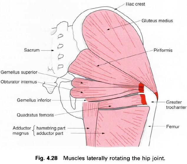
Piriformis
Obturator internus
Gemellus superior
Gemelus inferior
Quadratus femoris
Obturator externus
Piriformis
Piriformis is found posterior to the hip joint, being in the same plane as gluteus medius. It is triangular in
shape, with its base in the pelvis and apex in the gluteal region.
Its upper attachment is to the front of the second to fourth sacral
segments, coming between and lateral to the anterior sacral foramina. It
has an additional attachment from the gluteal
surface of the ilium and the pelvic surface of the sacrotuberous ligament as it passes out
of the pelvis through the greater sciatic foramen into the gluteal region. Its
fibres continue to pass downwards, laterally and forwards, narrowing into a
tendon which attaches to the upper border and medial side of the greater
trochanter of the femur. The
fibres run in a straight line from the origin to the insertion through the
greater sciatic foramen.
Nerve
supply
Piriformis is supplied from the anterior rami of the sacral plexus, L5,
S1, 2; and mainly S1. The skin covering this area is supplied by the same nerve
roots but it must be remembered that this is a deep muscle and the gluteus maximus intervenes between the
two.
Action
In the anatomical position this muscle will
certainly be a lateral rotator of the thigh, although it is situated a little
high. However, in the sitting position it is concerned in the important action
of the abduction, and in this particular situation is well positioned for this
action. It is an important muscle in holding the head of the femur in the acetabulum.
Functional
activity
It is well to remember that many times we only
consider the action of a muscle in the anatomical position. In the sitting
position, the pull of muscles changes and the movement produced may bear little
or no relation to the previous action. Piriformis will be particularly
concerned in the following activities:
- abduction when sitting, for example in moving from one chair to another without standing up;
- moving the legs to the outside of a car in preparation for standing up;
- stabilizing the pelvis when the trunk is rotated;
- controlling the balance of the pelvis when standing on a moving bus.
It is not therefore surprising that strain of
this muscle is quite common, but unfortunately easily overlooked.
Palpation
This is not easy as piriformis is situated deep
inside the buttock. Dig your fingers into the buttock just lateral to the sacrum and then push the outside of
the thigh up against the leg of a table or some such resistance; you can feel
then the muscle contract, although it must be remembered that a large part of
the muscle is in the pelvis.
Obturator internus
Obturator internus is again a triangularly
shaped muscle, situated partly in the pelvis and partly in the gluteal region
posterior to the hip joint. It arises
from the internal surface of the obturator membrane and surrounding bony
margin, except at the obturator canal. The bony attachment extends backwards as
far as the pelvic surface of the ilium. The muscle fibres pass laterally,
but mainly backwards towards the lesser sciatic foramen, through which they
pass, narrowing down and becoming tendinous.
As the tendon passes through the lesser sciatic
foramen deep to the sacrotuberous ligament it changes direction to the
sacrotuberous ligament it changes direction to pass forwards and laterally to
insert into the medial surface of the
greater trochanter of the femur in front of and above the
trochanteric fossa. Before attaching to the femur,
the tendon is commonly joined by the tendons of the gemellus superior above,
and the gemellus inferior below. Occasionally these two tendons will insert
into the greater trochanter above and below the tendon of the obturator
internus.
The inner(pelvic) surface of obturator internus
is covered by the obturator fascia, from which arises part of levator ani. As
the tendon of obturator internus turns around the lesser sciatic notch the
surface of the bone in this region is grooved and covered with cartilage. A
bursa intervenes between tendon and cartilage.
Nerve
supply
This muscle is supplied by the nerve to obturator internus, root value
L5, S1, 2. The skin covering this area is mainly supplied by S3.
Action
In the anatomical position this muscle is a
lateral rotator of the thigh pulling the greater trochanter backwards using the
hip joint as the fulcrum. However,
when the hip is flexed to a right
angle, it will pull the upper end of the femur
medially, and therefore the lower end will move laterally as in abduction.
Functional
activity
As in the case of the piriformis, obturator
internus will be used when moving sideways in the seated position, in swinging
the lower limb sideways, as in placing the limb outside a car, and in balancing
and controlling the stability of the trunk when the seated person is being
rocked from side to side. For the same reasons, moving around on the floor or
on a platform, either sitting or crawling will require considerable activity in
this muscle.
Gemellus superior
As obturator internus passes out of the pelvis
around the lesser sciatic notch it is joined by gemellus superior and inferior.
Gemellus superior arises from te gluteal surface of the ischial spine. It runs laterally and
slightly downwards to blend with the
superior aspect of the tendon of the obturator internus. Sometimes its
fibres are prolonged onto the medial surface of the greater trochanter of the femur.
Nerve
supply
The muscle is supplied by the nerve to obturator internus, root value L5, S1, 2.
Gemellus inferior
Gemellus inferior arises from the upper part of the ischial tuberosity. It runs laterally and slightly upwards to blend
with the inferior aspect of the tendon of obturator internus.
Nerve
supply
The muscle is supplied by the nerve to quadratus femoris, root value
L4, 5, S1.
Action
The gemelli aid obturator internus in its
action. As obturator internus turns around the lesser sciatic notch it will
lose some of its power; this will obviously be compensated for by the pull of
the gemelli.
Quadratus femoris
This muscle is situated below gemellus inferior
and above the upper margin of adductor magnus. It is a flat quadrilateral muscle, separated from the hip joint by the obturator externus.
It attaches to the ischial tuberosity just below the lower rim of the acetabulum. The
fibres pass laterally to attach to the quadrate
tubercle situated half way down the intertrochanteric crest of femur and the area of bone surrounding
it.
Nerve
supply
The muscle is supplied by the nerve to quadratus femoris, root value L4, 5, S1.
Action
In the anatomical position quadratus femoris is
obviously a lateral rotator of the hip
joint, but with the hip flexed, it
will act as an abductor of the hip.
Obturator externus
This is a triangular muscle having its muscular
base attached to the outer surface of
the obturator membrane and the
surrounding margins of the pubis and ischium, excluding the area superiorly
around the obturator canal. The muscle fibres converge on to a tendon which
runs in a groove below the acetabulum across the back of the neck of the femur, which it grooves, to insert into
the trochanteric fossa of femur. The
muscle lies deep to quadratus femoris.
Nerve
supply
The muscle is supplied by the posterior branch of the obturator nerve, root value L3, 4.
Action
In the anatomical position obturator externus
laterally rotates the femur. However,
when the hip is flexed it will pull
the upper part of the femur medially
with the lower part passing laterally, as in abduction.
Functional
activity
The functional activity of these muscles must
be considered together. Piriformis, the gemelli, obturator externus and
internus, and quadratus femoris are always considered in the anatomical
position. In this position these muscles will perform an important role in the
control of the pelvis, particularly when only one foot is on the ground and even more so in the walking. They are
responsible, together with gluteus maximus and the posterior part of gluteus medius, in producing lateral rotation of the lower limb in the forward
swing-through phase of gait. In sitting, crawling and turning over when lying
down they will, however, have a completely different role, producing abduction
of the hip and thereby controlling
the movements of the pelvis on the flexed thigh.
Palpation
These muscles are situated deep to the thick gluteus maximus and it is almost
impossible to distinguish the contraction of these muscles through the
overlying muscle tissue, especially as gluteus maximus is usually contracting at the same time. However, overactivity or
strain of these muscles may result in acute tenderness deep to the back of the hip joint; the production of this
pain with the relevant movement is then obvious.
It is difficult to research the actions of
these lateral rotators primarily because of their depth in the gluteal region,
but also because much of their action is concerned with the control of the
movements of the hip and pelvis. Thus
they may be in a state of contraction even when the opposite movement to their primary
action is occurring.
To see these muscles, gluteus maximus must be
removed – the muscles resembling the rungs of a ladder: consequently, they are
often referred to as the ladder of
muscles.


0 коментара:
Постави коментар