


The radial nerve
The radial nerve is the major nerve from the
posterior cord, root value C5, 6, 7, 8 (T1), being one of its terminal
branches. In the axilla, the radial nerve lies behind the axillary and upper
part of the brachial arteries, passing anterior to the tendons of subscapularis, latissimus dorsi and teres major. The radial nerve, together with the profunda brachii artery, enters
the posterior compartment of the arm by passing through the lower triangular
space, formed by the humerus
laterally, the long head of triceps
medially and teres major above. In
passing through this space, the nerve enters the spiral(or radial) groove of
the humerus, descending obliquely
between the lateral border of the humerus in the distal third of the arm. The nerve pierces the lateral and medial
heads of triceps, reaching the
lateral border of the humerus in the
distal third of the arm. The nerve pierces the lateral intermuscular septum to
enter the anterior compartment where it lies in a muscular groove between brachialis and brachioradialis. In front
of the lateral epicondyle of the humerus,
the radial nerve divides into its terminal superficial and deep branches.
In the arm, the radial nerve gives a supply to
all three heads of triceps, anconeus,
the lateral part of brachialis, brachioradialis and extensor carpi radialis longus. The branches to triceps
all arise before the radial nerve enters the spiral groove; anconeus is supplied by a branch to the
medial head of triceps.
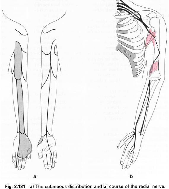
The radial nerve also gives articular branches
to the elbow joint and has three cutaneous branches which supply skin on the
back of the arm and forearm(figure a).
The
posterior cutaneous nerve of
the arm arises in the axilla, piercing the deep fascia near the posterior
axillary fold. It supplies skin on the posterior surface of the proximal third
of the arm.
The
lower cutaneous nerve of the arm
arises before the radial nerve pierces the lateral intermuscular septum, and
becomes cutaneous just below deltoid.
It supplies the skin over the lower lateral part of the arm and a small area on
the forearm.
The
posterior cutaneous nerve of the forearm arises just below the previous nerve, and supplies a variable area of
skin on the dorsum of the forearm as far as the wrist, or occasionally beyond.
The
superficial branch is the
direct continuation of the radial nerve, beginning in front of the lateral
epicondyle and descending along the anterolateral side of the forearm. It is
entirely sensory. It lies on supinator,
pronator teres, flexor digitorum superficialis and flexor pollicis longus covered by brachioradialis with the radial artery medial to it. In the distal
third of the forearm, the nerve passes below the tendon of brachioradialis and pierces the deep fascia to become superficial.
It supplies the skin on the dorsum of the wrist, the lateral dorsal surface of
the hand and dorsum of the thumb, and
then divides into four or five digital nerves. The digital nerves supply the
skin on the dorsum of the thumb, index, middle and adjacent half of the ring
finger as far as the distal interphalangeal joint. The digital branches also
give articular branches to the metacarpophalangeal and proximal interphalangeal
joints of all five digits.
The deep
branch, more often called the posterior
interosseus nerve, is entirely muscular and articular. It begins in front
of the lateral epicondyle of the humerus and enters the posterior compartment of the forearm by passing between the two
heads of supinator, thereby curving
around the lateral and posterior surfaces of the radius. During its course, the nerve supplies both extensor carpi radialis brevis and supinator. It then descends between the
deep and superficial groups of extensor muscles, accompanied by the posterior
interosseus artery, supplying all the muscles in the extensor compartment of
the forearm: extensor digitorum, extensordigiti minimi, extensor carpi ulnaris, extensor pollicis longus, extensorindicis, abductor pollicis longus and extensor pollicis brevis.
In the lower part of the forearm, the posterior interosseus nerve lies on the
interosseus membrane and ends in a flattened expansion, which gives articular
branches to the intercarpal joints.
Applied
anatomy
The radial nerve is often injured as it crosses
the humerus, either as the result of
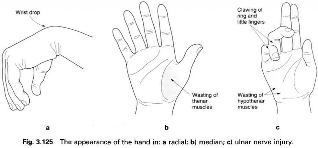
a fracture or by pressure from a direct blow or incorrect use of a crutch. Triceps usually escapes denervation as
it derives its supply from branches given off high in the arm, but a total
paralysis of the extensors of the wrist
and digits leads to the deformity of a “dropped wrist”(figure a). As a
result, any attempt to grip or make a fist leads to increased flexion of the
wrist and an inability to carry out effective movement. This is due to the loss
of the synergic action of the wrist extensors which usually prevent the
unwanted flexion of the wrist produced by the continued action of the finger flexors.
The interphalangeal joints of the fingers can
be extended by the lumbricals and interossei which have an attachment to
the dorsal digital expansion, but proper use of the hand requires effective form of “lively” splint which compensates
for the paralysed muscles. Even though the sensory distribution of the radial
nerve on the dorsum of the hand appears
extensive, overlap by adjacent cutaneous nerves means that the area of
exclusive radial nerve supply is a small patch on the dorsum of the thumb web.
The median nerve
The median nerve is complex in that it arises
partly from the lateral cord (C5, 6, 7) and partly from the medial cord (C8,
T1) of the brachial plexus. These two contributing heads of the median nerve
unite by embracing the third part of the axillary artery. Once formed, the
nerve descends under cover of biceps
passing at first laterally to the brachial artery and then medially, having
crossed it anteriorly. In the lower part of the arm the median nerve lies on brachialis, and in the cubital fossa is
protected by the bicipital aponeurosis which crosses it.
The median nerve enters the forearm by passing
between the two heads of pronator teres,
and then runs below the tendinous arch connecting the heads of flexor digitorum superficialis to gain
access to its deep surface. Closely bound to the deep surface of flexor digitorum superficialis, it
descends on flexor digitorum profundus
until just above the wrist where it becomes superficial by passing between the
tendons of flexor digitorum superficialis and flexor carpi radialis, deep to palmaris longus. The median nerve enters the hand
deep to the flexor retinaculum, passing anteriorly to the long flexor tendons. Consequently,
it is one fo the structures found within the carpal tunnel.
During its course the median nerve gives
articular branches to the elbow joint and supplies pronator teres, flexor carpi radialis, palmaris longus and flexor digitorum superficialis.
The palmar
cutaneous nerve arises in the distal third of the forearm. It pierces the
deep fascia and enters the palm by passing superficial to the flexor
retinaculum. It supplies a small area of skin on the lateral side of the palm
and thenar eminence.
In the cubital fossa, the anterior interosseus nerve arises from the median nerve and
descends, with the anterior interosseus artery, on the anterior surface of the
interosseus membrane between flexor pollicis longus and flexor digitorum profundus. It then runs deep to pronator quadratus eo end at the wrist
by giving articular branches to the radiocarpal and intercarpal joints. The
anterior interosseus nerve supplies flexor pollicis longus, the lateral half of flexor digitorum profundus and pronator quadratus.
Once the median nerve has passed through the
carpal tunnel to enter the hand, it
divides into lateral and medial terminal branches. The lateral branch passes laterally and proximally to enter the thenar
eminence and supply abductor pollicis brevis, flexor pollicis brevis, opponens pollicis and the first lumbrical. It gives sensory branches to
the adjacent sides of the thumb and index finger.
The medial
branch of the median nerve divides into a variable number of branches, the
palmar digital nerves, the most lateral of which supplies the second lumbrical.
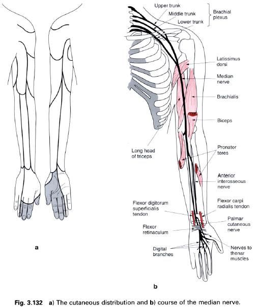
These nerves are sensory to the palmar surface of adjacent sides of the index
and middle, and middle and ring fingers(figure a). Each of these digital nerves
produces a dorsal branch which passes backwards to supply the dorsal aspect of
the distal phalanx and nail bed, and a variable amount of the middle phalanx of
the same digits.
The digital
nerves lie deep to the palmar aponeurosis and superficial palmar arch, but
superficial to the long flexor tendons. As well as the sensory innervation,
they also give articular branches to the interphalangeal and
metacarpophalangeal joints.
Applied
anatomy
The median nerve can be injured in the forearm
by deep cuts with a resultant loss of flexion at all interphalangeal joints,
except the distal ones in the ring and little fingers. The metacarpophalangeal
joints of these same fingers can still be flexed by the lumbricals and interossei
but the movement of pronation is severely restricted. In the hand, the thumb is held in extension and
adduction thus losing its ability to oppose and abduct. This, combined with the
sensory loss, proves a major disability. More commonly the nerve is damaged
just proximal to the flexor retinaculum by laceration, or deep to it in the
carpal tunnel where compression gives rise to the carpal tunnel syndrome. In
this instance only the thenar muscles, lateral two lumbricals and sensation in
the hand will be affected.


0 коментара:
Постави коментар