Free Facebook Likes, Youtube Subscribers,  Twitter Followers

Ads 468x60px

Blogger Tricks

Blogger Themes

Приказивање постова са ознаком Anatomy. Прикажи све постове

4. 8. 2013.

Muscles adducting the toes


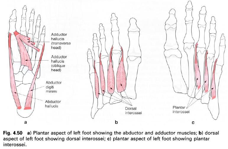
Adductor hallucis

Adductor hallucis is situated deep within the plantar aspect of the foot. It arises by two heads, oblique and transverse. The oblique head comes from the plantar surface of the based of the second, third and fourth metatarsals and the sheath of the tendon of peroneus longus. The transverse head comes from the plantar surface of the lateral three metatarsophalangeal joints and the deep transverse metatarsal ligament.
The muscle fibers of the oblique head pass forwards and medially while those of the transverse head pass medially. The two heads unite and blend with the medial part of the flexor hallucis brevis to insert into the lateral side of the base of the proximal phalanx of the great toe.

Nerve supply

Adductor hallucis is supplied by the lateral plantar nerve, root value S2, 3, and the skin covering this area is supplied by root S1.


Action

It will adduct the great toe towards the second toe, and flex the first metatarsophalangeal joint.

Functional activity

Working with abductor hallucis, adductor hallucis helps to control the position of the great toe so that active flexion can be produced and thereby provide the final thrust needed in walking, running or jumping. Due to its transverse position across the forefoot it will also help to maintain the anterior metatarsal arch of the foot.
The pull of adductor hallucis is almost at right angles to the phalanx and therefore will have a better mechanical advantage than abductor hallucis. If the medial longitudinal arch is allowed to fall, allowing the foot to drift medially and the toes laterally, the pull of adductor hallucis will overcome that of the abductor, thus adding to the deformity often seen in the great toe.

Palpation

This muscle is too deep to be palpated.

Muscles abducting the toes


Abductor hallucis
Abductor digiti minimi

Abductor hallucis

Abductor hallucis is a powerful and important muscle found superficially on the medial side of the plantar aspect of the foot, lying deep to the medial part of the plantar aponeurosis. It arises, in part, from the plantar aponeurosis, the plantar aspect of the medial tubercle of the calcaneus, the flexor retinaculum and the intermuscular septum separating it from flexor digitorum brevis.
The fibres pass forwards forming a tendon which passes over the medial side of the metatarsophalangeal joint of the great toe, to insert into the medial side of he base of the proximal phalanx in conjunction with the tendon of the flexor hallucis brevis.

Nerve supply

Abductor hallucis is supplied by the medial plantar nerve, root value S1, 2, with the skin covering the muscle supplied by root L5.


Action

As its name implies, the muscle abducts the great toe at the metatarsophalangeal joint and also helps to flex it at this point.

Functional activity

Abduction of the great toe is not of importance as such, except perhaps as a party trick and then very few people are able to perform the action easily! However, the muscle is strong and bulky, and it must therefore be assumed that it has an important role to play in some specific activity.
Due to its position along the medial side of the foot, together with the fact that it is attached at the back and front of the medial longitudinal arch, it can act as a bow-string to the arch when the foot is being used for propelling the body forwards. Its attachment to the medial side of the great toe also helps in controlling the central position of this toe when it is being flexed.
It should be noted that when the muscle contracts hard, the great toe does indeed move medially, but more importantly, the foot is positioned laterally, thus improving the relationship between great toe and medial side of the foot. Indeed, if this alignment of the foot and toes was encouraged from an early age many deformities of the toes may be prevented.

Palpation

Place the fingers on the medial plantar aspect of the foot, under the medial longitudinal arch. The toes are then flexed and the belly of the muscle can be easily palpated towards the heel. Tracing forwards from the heel, the tendon of the muscle can be felt.

Abductor digiti minimi

Abductor digiti minimi is found on the lateral side of the plantar aspect of the foot, lying deep to the plantar aponeurosis from which it gains part of its attachment. It also arises from the medial and lateral tubercles of the calcaneus and from the area between, as well as the intermuscular septum is separating it from flexor digitorum brevis.
The fibres pass forwards forming a tendon which inserts into the lateral side of the base of the proximal phalanx of the fifth toe.

Nerve supply

Abductor digiti minimi is supplied by the lateral plantar nerve, root value S2, 3, with the skin covering the muscle being supplied by root S1.

Action

On contraction abductor digiti minimi abducts the fifth toe at the metatarsophalangeal joint and also helps to flex it at this joint.

Functional activity

Because the muscle runs from the posterior to the anterior parts of the lateral longitudinal arch, it acts as a bow-string to this arch in a similar way to abductor hallucis on the medial side of the foot, except of course that the lateral arch can hardly be called a true arch. Nevertheless, the muscle certainly comes into action in running and jumping activities to ensure that this arch is maintained under stress.

Palpation

Unless a subject can abduct the fifth toe easily, the muscle is difficult to palpate.

Abduction and adduction of the toes


In the hand, it is the middle finger which is regarded as the central digit when considering abduction and adduction. In the foot, however, the central digit when considering these movements is the second toe. Therefore, if the great toe is drawn medially it is said to abduct, whereas if all the other toes are drawn laterally, i.e. away from the second toe, that is also termed abduction. If all the toes are drawn towards the second toe they are said to adduct.

21. 7. 2013.

Muscles flexing the toes


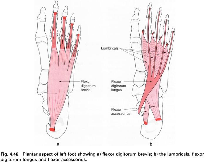
Flexor accessorius (quadratus plantae)
Flexor digitorum brevis
Flexor hallucis brevis

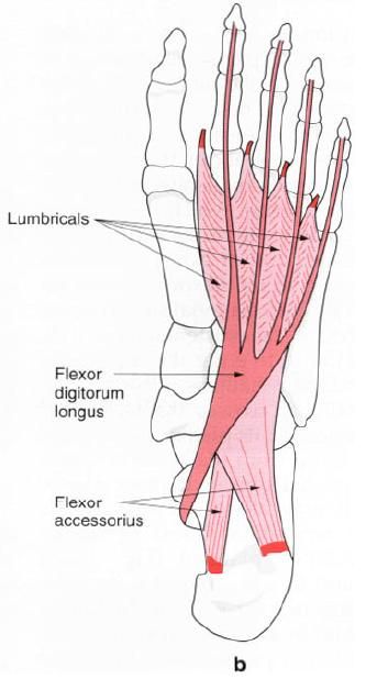
Flexor accessorius (quadratus plantae)

Flexor accessorius lies deep to flexor digitorum brevis, arising by two heads from the medial and lateral tubercles of the calcaneus and the adjacent long plantar ligament. A flattened muscular band is formed by the merging of the two heads which inserts into the tendon of flexor digitorum longus in the midpoint of the sole, proximal to the origin of the lumbricals.


Nerve supply

Flexor accessorius is supplied by the lateral plantar nerve, root value S2, 3. The skin over the region is supplied by root S1.

Action

It helps the long flexor tendons flex all the joints of the lateral four toes. By pulling on the lateral side of the tendon of flexor digitorum longus, it changes the direction of pull so that the toes flex towards the heel and not towards the medial malleolus.

Functional activity

Flexor accessorius has an important role to play in gait when flexor digitorum longus is already shortened because of plantarflexion of the ankle joint. This muscle exerts its action on the long flexor tendons so that the toes can be flexed to grip the ground giving support and thrust during the propulsive phase. This action essentially means that flexor digitorum longus can be considered to act powerfully across two joints at the same time – an unusual phenomenon.

Palpation

Lying deep in the sole of the foot, flexor accessorius cannot be palpated.

Flexor digitorum brevis

Flexor digitorum brevis is situated in the sole of the foot lying just deep to the central part of the plantar aponeurosis between abductor hallucis medially and abductor digiti minimi laterally. Arising from the medial tubercle of the calcaneus, the deep surface of the central portion of the plantar aponeurosis and the muscular septa either side, the fibres pass forwards in the middle of the sole, and separate into four tendons, which pass to the lateral four toes. Just distal to the metatarsophalangeal joint, within their respective fibrous flexor sheaths, each tendon splits into two for the passage of the flexor digitorum longus tendon, which passes from deep to superficial. After rotating through almost 180° the outer margins of the slips of each tendon rejoin, leaving a shallow groove along which the flexor digitorum longus tendon slides. After passing over the proximal interphalangeal joint, the tendon again splits to insert into the sides of the base of the middle phalanx.

Nerve supply

Flexor digitorum brevis is supplied by the medial plantar nerve, root value S2, 3. The skin covering this area is supplied by roots L5, S1.

Action

Flexor digitorum brevis primarily flexes the proximal interphalangeal joint of the lateral four toes, followed by flexion of the metatarsophalangeal joints.

Functional activity

Flexor digitorum brevis is obviously concerned, as is flexor digitorum longus, in producing the thrust from the toes when the demand arises.

Palpation

This muscle is almost impossible to palpate as it is covered with some of the thickest fascia in the body and its tendons lie deep within the foot.

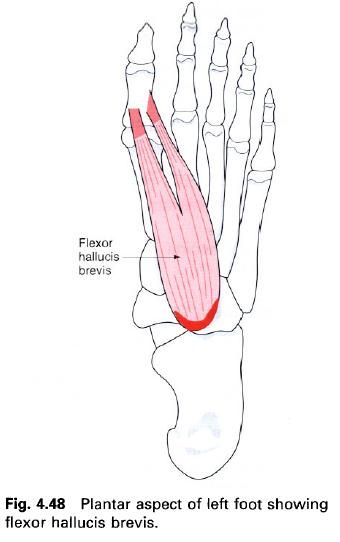
Flexor hallucis brevis

Flexor hallucis brevis is a short muscle situated deep in the sole of the foot, appearing between abductor hallucis medially and flexor digitorum brevis laterally. It arises from the medial side of the plantar surface of the cuboid bone, behind the groove for peroneus longus, and the adjacent surface of the lateral cuneiform and from the tendon of tibialis posterior.
The muscle fibres run forwards and medially towards the great toe, separating into two fleshy bellies to lie either side and deep to the tendon of flexor hallucis longus. From each belly arises a tendon which inserts onto the appropriate side of the base of the proximal phalanx. The medial tendon unites with that of abductor hallucis, while the lateral tendon unites with the tendon of adductor hallucis, thereby giving common insertions. Small sesamoid bones, which run in shallow grooves on the head of the first metatarsal, develop in each tendon.

Nerve supply

Flexor hallucis brevis is supplied by the medial plantar nerve, root value S1, 2. The skin covering the area is supplied by root L5.

Action

The action of flexor hallucis brevis is to flex the metatarsophalangeal joint of the great toe.


Functional activity

Flexor hallucis brevis will, of course, aid flexor hallucis longus in the final push-off from the ground during activity. Being accompanied at its insertion by abductor and adductor hallucis suggests that the steadying of the great toe during propulsion must be of great importance, probably to ensure the generation of maximum force. When the great toe is deformed as in hallux valgus, where the tip points laterally and the base medially, this thrust is lost and the patient finds it difficult to run or sometimes walk, even at slow speeds.
It is interesting to note that it is not uncommon for injuries to occur to the sesamoid bones, particularly in individuals who put considerable strain on the great toe. Such injuries produce an inflamed region where the sesamoid bone slides against the metatarsal. This can cause considerable pain and altered function.

Palpation

This muscle is set so deep in the plantar surface of the foot that it is not possible to palpate. Only the sesamoid bones found within its tendons can be felt, and then only with considerable practice.

29. 6. 2013.

Muscles extending the toes


Extensor digitorum brevis
The lumbricals
The interossei

Extensor digitorum brevis

Extensor digitorum brevis is situated on the dorsum of the foot beyond the inferior part of the extensor retinaculum, lateral to and partly covered by the tendons of peroneus tertius and extensor digitorum longus. It is a thin muscle arising from the anterior roughened part of the upper surface of the calcaneus and the deep fascia covering the muscle, including the stem of the inferior extensor retinaculum. From the small belly, short tendons pass forwards and medially, the most medial of which crosses the dorsalis pedis artery to insert separately on to the dorsal aspect of the base of the proximal phalanx of the great toe. The remaining three tendons join the lateral side of the dorsal hood of the second, third and fourth toes. The most medial part of the muscle may develop a separate belly, sometimes reffered to as extensor hallucis brevis.

Nerve supply

This muscle is supplied by the deep peroneal nerve, root value L5, S1. The skin covering the muscle is supplied by roots L5, S1.

Action

The medial part of the muscle aids extensor hallucis longus in extending the great toe at the metatarsophalangeal joint, while the other three tendons aid extensor digitorum longus. As with the long extensor tendons, extensor digitorum brevis helps the lumbricals to extend the interphalangeal joints; however, it is unable to do this independently.

Functional activity

Extensor digitorum brevis will help extensor digitorum longus and extensor hallucis longus to raise the toes clear to the ground in running and walking.

Palpation

Place the fingers on the tendon of extensor digitorum longus as it splits into its four parts. When the toes are extended, extensor digitorum brevis can be felt just lateral and deep to the tendon. The tendons are difficult to trace distally as they become inseparable from those of extensor digitorum longus.

The lumbricals

These are four small muscles associated with the tendons of flexor digitorum longus; they pass from the flexor to the extensor compartment of the foot. The most medial of these muscles arises from the medial side of the tendon to the second toe, adjacent to the attachment of flexor accessories (quadratus plantae) to the main longus tendon. The remaining lumbricals arise by two heads from adjacent sides of two tendons, that is the second from the tendons to the second and third toes, the third from the tendons to the third and fourth toes, and the fourth from the tendons to the fourth and fifth toes. Each muscle then passes forwards below the deep transverse metatarsal ligament on the medial side of the toe, winding obliquely upwards to attach to the medial side of the extensor hood and base of the proximal phalanx.

Nerve supply

The first and most medial lumbrical is supplied by the medial plantar nerve, root value S1, 2, while the lateral three are supplied by the lateral plantar nerve, root value S2, 3; both being terminal branches of the tibial nerve. The skin on the dorsum of the foot at the point of attachment is supplied by roots L5, S1. The skin of the plantar aspect of the foot overlying the muscles is supplied by the medial and lateral plantar nerves, which have the same root values as the supply to the muscles. It should be noted, however, that only the most lateral of the lumbricals has skin over its plantar aspect.


Action

There has been much discussion about the role of these small, almost insignificant muscles. They have a long muscle belly compared with their tendon and they link the flexors of the toes with the extensors. By their attachment to the proximal phalanx, contraction of the lumbricals produces flexion of the toes at the metatarsophalangeal joint. However, because they also insert into the extensor hood, the lumbricals extend the interphalangeal joints. Indeed, this latter action is primarily due to the lumbricals and not the long and short extensor tendons.

Functional activity

The action of the lumbricals prevents clawing of the toes in the propulsive phase of gait. Paralysis of these muscles results in the extensor muscles pulling the toes into hyperextension at the metatarsophalangeal joint. Even at rest the toes become clawed.
The nerves which supply these muscles appear to have many more fibres than would be necessary for such a small muscle and a great number of these are sensory. This leads one to believe that they may have a very important function in providing information related to the tension developed between the long flexor and extensor muscles. This sort of information is of great importance in locomotion, especially as the point of attachment of the lumbricals is a long way from the muscle bellies of the extensors and flexors.

Palpation

It is not possible to palpate these muscles as they lie deep in the sole of the foot covered by many of the small muscles of the sole and the long flexor tendons.

Dorsal interossei

There are four dorsal interossei, being small bipennate muscles situated between the metatarsals. Each arises from the proximal half of the sides of adjacent metatarsals, forming a central tendon which passes forwards, deep to the deep transverse metatarsal ligament. It passes between the metatarsal heads to attach to the side of the proximal phalanx and capsule of the metatarsophalangeal joint. The tendons do not attach to the extensor hood.
The first, or most medial, arises from the adjacent sides of the first and second metatarsals and attaches to the medial side of the base of the proximal phalanx of the second toe. The second arises from the adjacent sides of the second and third metatarsals and it also attaches to the proximal phalanx of the second toe but to the lateral side. The third and fourth  dorsal interossei attach to the lateral side of the proximal phalanx of the third and fourth toes respectively.

Nerve supply

All four dorsal interossei are supplied by the lateral plantar nerve, root value S2, 3, those in the fourth interosseus space from the superficial branch, and the rest by the deep branch. The skin covering this area on the dorsum of the foot is supplied by root L5 medially and S1 laterally.

Action

The dorsal interossei abduct the toes at the metatarsophalangeal joint, however this action, as such, is of little importance in the foot. Acting with the plantar interossei, they will produce flexion of the metatarsophalangeal joint.

Functional activity

The dorsal interossei are powerful little muscles and their activity in combination with the plantar interossei controls the direction of the toes during violent activity, thus enabling the long and short flexors to perform their appropriate actions.
These muscles, because of their relationship to the metatarsophalangeal joint, can flex these joints and so raise the heads of the second, third and fourth metatarsals, thus helping to maintain the anterior metatarsal arch. They also help, to a limited extent, with the maintenance of the medial and lateral longitudinal arches of the foot.

Palpation

Place the finger tips between the proximal parts of the metatarsals on the dorsum of the foot; when the toes are abducted; the muscles can be felt to contract.


Plantar interossei

The plantar interossei are smaller than their dorsal counterparts, fusiform in shape and found in the lateral three interosseus spaces. Each arises from the plantar and medial aspect of the base and proximal end of the shaft of the metatarsal. The tendon formed passes forwards and deep to the deep transverse metatarsal ligament to insert into the medial side of the base of the proximal phalanx of the same toe.

Nerve supply

All the interossei are supplied by the lateral plantar nerve, root value S2, 3, with that in the fourth interosseus space being supplied by the superficial branch of the nerve. The skin covering the area is supplied on the lateral side by root S1 and medially by root L5.

Action

The plantar interossei adduct the third, fourth and fifth toes towards the second. In conjunction with the dorsal interossei they flex the metatarsophalangeal joints of the lateral three toes.

Functional activity

With the help of the dorsal interossei and abductor digiti minimi, the plantar interossei help to control the position of the third, fourth and fifth toes during the push-off phase of walking and running. They also help to prevent splaying of the toes when weight is suddenly applied to the forefoot.

Palpation

These muscles are too deep to be palpated. 

3. 6. 2013.

Muscles everting the foot


Peroneus brevis

Peroneus brevis

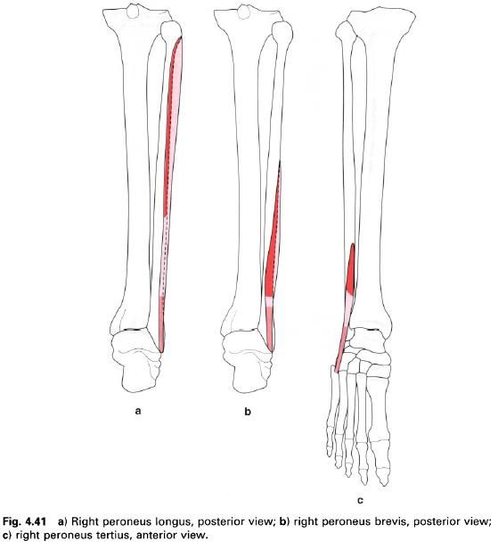
Peroneus brevis is also situated on the lateral side of the leg enclosed in the same osteofascial compartment. It arises from the lower two-thirds of the lateral surface of the fibula, the upper half being anterior to peroneus longus. It also attaches to the intermuscular septa at all sides.
The muscle belly is fusiform and short, soon passing into a tendon which accompanies that of peroneus longus to pass behind the lateral malleolus in a common synovial sheath. The tendon then passes forwards and downwards into a groove above the peroneal tubercle of the calcaneus, which is converted into a tunnel by the inferior part of the peroneal retinaculum. It then passes forward to its insertion into the tubercle on the lateral side of the base of the fifth metatarsal. Above the tubercle, the tendon is surrounded by a synovial sheath, which is separated from that of peroneus longus. A slip from the tendon usually joins the long extensor to the little toe. Other separate slips may join peroneus longus, or pass to the calcaneus or cuboid.

Nerve supply

Peroneus brevis is supplied by the superficial peroneal nerve, root value L5, S1. The skin covering the muscle is innervated by L5, S1, 2.

Action

Peroneus brevis is an evertor of the foot. Because of its course and attachments, the pull of its tendon is in such a direction to produce plantarflexion of the ankle at the same time. 


Functional activity

This muscle is also well-positioned to prevent sideways sway in the standing position. In standing on one leg, it will help to prevent the body falling to the opposite side, thus working with a reversed origin and insertion. In walking or running, especially over the rough ground, it plays an important role in controlling the position of the foot and should prevent the foot from becoming too inverted. In many cases, however, this mechanism does not always appear to work correctly, and the foot over-inverts causing the weight to come down on the lateral side of the foot forcing the foot into further inversion. This can severely damage or even snap the tendon of the muscle and often the anterior talofibular ligament of the ankle joint.

Palpation

If the fingers are placed on the belly of peroneus longus and then moved downwards to the lower half of the fibula (but in the same vertical line), the belly of peroneus brevis can be palpated when the foot is everted and plantarflexed. Its tendon can easily be traced to the groove just above the peroneal tubercle and then forwards to its insertion into the tubercle of the fifth metatarsal.


29. 5. 2013.

Muscles inverting the foot

Muscles dorsiflexing the ankle


Tibialis anterior
Extensor digitorum longus
Extensor hallucis longus
Peroneus tertius

Tibialis anterior

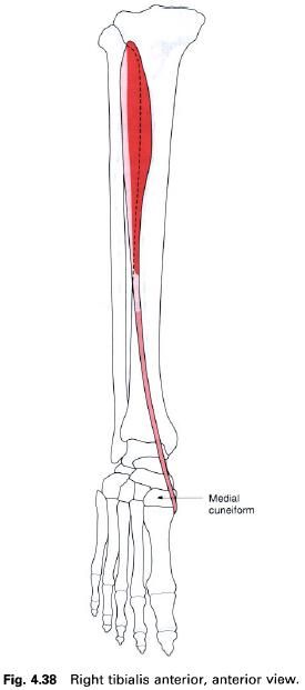
Tibialis anterior is a long fusiform muscle situated on the front of the leg lateral to the anterior border of the tibia. It is covered by strong fascia and gains its upper attachment from the deep surface of this fascia, the upper two-thirds of the lateral surface of the tibia and the adjoining part of the interosseus membrane. The muscle becomes tendinous in its lower third, passing downwards and medially over the distal end of the tibia. The tendon continues through both the superior and inferior extensor retinaculae to insert into the medial side of the medial cuneiform and base of the first metatarsal, the insertion reaching the under surface of both bones to blend with that of peroneus longus.

Nerve supply

This muscle is supplied by the deep peroneal nerve, root value L4, 5. The skin covering the muscle is also supplied by roots L4, 5.

Action

Tibialis anterior is a dorsiflexor of the foot at the ankle joint. When working with tibialis posterior it acts as an invertor of the foot, in which the sole of the foot is turned to face medially.


Functional activity

As with other muscles in the leg, tibialis anterior is concerned with balancing the body on the foot. It works with the surrounding muscles to maintain body balance during activities of the upper part of the body which change the distribution of weight.
Not only is tibialis anterior responsible for dorsiflexing the foot as the lower limb is carried forward during the swing-through phase of walking, so preventing the toes catching the ground, it also controls the placement of the foot on the ground following initial ground contact by the heel. On close observation, especially in slow motion, it will be seen that the heel does not strike the ground and remain immobile at the initiation of the stance phase, but glides on to the surface and acts as the first braking force of the lower limb’s forward movement. Overactivity of tibialis anterior accounts for the wear patterns seen on the posterolateral aspect of the heel, due to the frictional forces between the shoe and the ground. The rest of the foot is then gradually lowered to the ground in a controlled manner taking up the undulations of the surface concerned. The landing of the foot on the ground is similar to the landing of an aeroplane; the main wheels touch down first applying the initial braking force followed by a controlled lowering of the front of the craft as the speed decreases.
Tibialis anterior in association with the other dorsiflexors, therefore, plays an important part in the lowering of the forefoot to the ground in walking or running and will be put under stress in extended activity particularly over rough terrain. The anterior calf muscles are enclosed in particularly tight fascia which allows very little expansion of the tissues. The result is a compression of the muscle during activity and a dragging on the attachments of the surrounding fascia, particularly where it attaches to the bone. This leads to a painful condition of this area commonly called “shin splints”.
Paralysis of tibialis anterior causes footdrop because the remaining dorsiflexors are not strong enough to raise the toes and so prevent them dragging along the ground. The patient may overcome this by flexing the knee more than normal during walking or by fitting a “toe-raise” orthosis to patients or their shoe.

Palpation

Both the muscle belly and tendon can be seen and felt when the foot is dorsiflexed against resistance, the tendon being the most medial at the ankle joint.

Extensor digitorum longus

Extensor digitorum longus is again situated on the anterior aspect of the leg, being lateral to tibialis anterior, and overlying extensor hallucis longus. It has a linear origin from the upper-two thirds of the anterior surface of the fibula, the deep fascia and the upper part of the interosseus membrane with its upper fibres reaching across the lateral condyle of the tibia in conjunction with those of peroneus longus. It is a pennate muscle with the tendon appearing on the medial side; the muscle fibres pass downwards and medially to reach it. The tendon passes over the front of the ankle joint deep to the superior extensor retinaculum and then through the loop of inferior extensor retinaculum accompanied by peroneus tertius. At the level of the inferior extensor retinaculum or immediately distal, it gives rise to four tendons which run to the lateral four toes. The four separate tendons are enclosed in a common synovial sheath at the level of the inferior extensor retinaculum. On the dorsal surface of the proximal phalanx, each tendon forms a triangular membranous expansion, known as the extensor hood (dorsal digital expansion). Each hood is joined on its medial side by the tendon of the lumbrical and on the lateral side for the second to fourth toes by the tendon of extensor digitorum brevis. The interossei of the foot do not have an attachment to the extensor hood.
As the hood passes forwards over the proximal phalanx it divides into three parts before reaching the dorsum of the proximal interphalangeal joint. The central portion attaches to the base of the middle phalanx, while the two outer portions unite before inserting on to the base of the distal phalanx. An attachment of the extensor hood to the dorsal aspect of the proximal phalanx has also been described.

Nerve supply

This muscle is supplied by the deep peroneal nerve, root value L5, S1. The skin covering the muscle is supplied by root L5.


Action

As its name implies, extensor digitorum longs is an extensor of the lateral four toes at the metatarsophalangeal joints, and also assists in extension at the interphalangeal joints. However it is unable to perform the latter action unaided, which is primarily performed by the lumbricals. If the lumbricals are paralysed, extensor digitorum longus produces hyperextension of the metatarsophalangeal joint, while the interphalangeal joints become flexed. As the muscle passes across the front of the ankle joint, it also aids in dorsiflexion of the foot.

Functional activity

During walking and running extensor digitorum longus draws the toes upwards after they have been flexed prior to toe-off, and keeps them clear of the ground until the heel and foot make contact with the ground again. Unfortunately, the lateral four toes in most individuals tend to be flexed at the proximal interphalangeal joint and extended at the distal interphalangeal joint. Consequently extensor digitorum longus will lift the toes in this adapted position.

Palpation

The muscle belly is easily palpated on the anterolateral aspect of the leg. From the head of the fibula on the lateral side of the leg, just below the knee joint, run the fingers downwards and medially for about 2cm. When raising the toes off the floor, the muscle can be felt contracting. Now place the fingers over the front of the ankle joint; the tendon can be identified standing out clearly, being lateral to those of tibialis anterior and extensor hallucis longus. From here the tendon can now either be traced upwards, under the superior part of the extensor retinaculum to join the muscle belly, or downwards where it breaks up into four individual tendons running towards each of the lateral four toes. Each tendon stands clear of the metatarsophalangeal joint as it passes towards the dorsum of the toe.

Extensor hallucis longus

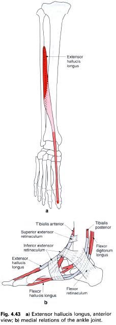
Extensor hallucis longus is situated deep to and between tibialis anterior and extensor digitorum longus on the front of the leg. Arising from the middle half of the anterior surface of the fibula and the adjacent interosseus membrane, the muscle fibres pass downwards and medially to the tendon which forms on its anterior surface. In this respect it is a unipennate muscle. The tendon passes under the superior extensor retinaculum, through the upper part of the inferior extensor retinaculum in a separate compartment enclosed in its own synovial sheath, and then deep to the lower band of the inferior extensor retinaculum on its way towards the base of the great toe. Generally, the tendon does not form a fully developed extensor hood but passes to attach to the base of the distal phalanx on its dorsal surface. Tendinous slips may be given off to the dorsal aspect of the base of the proximal phalanx and the first metatarsal.

Nerve supply

Extensor hallucis longus is supplied by the deep peroneal nerve, root value L5, S1. The skin covering this area is supplied by roots L4, 5.

Action

As its name implies, extensor hallucis longus will extend all of the joints of the great toe, but mainly the metatarsophalangeal joint. It is also a powerful dorsiflexor of the foot at the ankle joint.


Functional activity

In running, the great toe is the last part of the foot to leave the ground and therefore the final thrust will come from the long flexors of the toes. After this, the toe must be brought back into the extended position at the same time as the foot is dorsiflexed and slightly inverted, ready for the heel to be placed on the ground for the next weightbearing phase. By extending the great toe and dorsiflexing the foot, clearance of the surface is also achieved. It should be noted that the great toe does not have a lumbrical muscle or interossei associated with it. Consequently, extension of the interphalangeal joint depends entirely on extensor hallucis longus. Paralysis of the muscle will result in flexion of the joint and buckling of the toe during the last phase of gait, due to the unopposed action of the flexor muscles.

Palpation

If the great toe is extended, the tendon of the muscle is clearly visible as it crosses the first metatarsophalangeal joint to its insertion into the base of the distal phalanx. Trace the fingers up the tendon; it can be felt and seen crossing the anterior aspect of the ankle joint lateral to the tendon of tibialis anterior. From here the tendon can be felt passing upwards and laterally before passing deep to the surrounding muscles. Continue to move the fingers upwards for another 12cm and allow them pass a little laterally; when the great toe is rhythmically extended and flexed, the muscle can just be felt contracting under the fingers.

Peroneus tertius

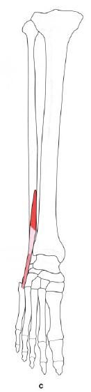
Peroneus tertius is situated on the lower lateral aspect of the leg and appears to have been part of extensor digitorum longus. It arises from the front of the lower quarter of the fibula in continuation with the attachment of extensor digitorum longus (with no gap between them), and from the intermuscular septum and adjoining fascia. Its fibres pass downwards and laterally into a tendon which passes deep to the superior and through the inferior extensor retinacula to insert into the medial and dorsal aspect of the base of the fifth metatarsal.

Nerve supply

Peroneus tertius is supplied by the deep peroneal nerve, root value L5, S1. The area of skin covering the muscle is also supplied by roots L5, S1.


Action

The muscle acts as a weak evertor and dorsiflexor of the foot at the ankle joint.

Functional activity

It is difficult to assess the importance of this small muscle as its actions appear to be covered by other muscles which have a much better mechanical leverage. Indeed in some subjects it is absent. It does, however, pass over the anterior talofibular ligament of the ankle joint, and it is well-known that this is very often damaged in inversion injuries. It is therefore well placed to help prevent too much inversion during sports activities, for example, and may be responsible for keeping down the number of injuries. Unfortunately, the muscle is often torn and may be completely ruptured during violent inversion, which is the cause of considerable pain and swelling. It is possible that with the attainment of bidepalism, peroneus tertius is assuming a more important role because eversion of the foot is a peculiarly human characteristic.

Palpation

Peroneus tertius is very difficult to palpate. However, it can be felt by drawing the fingers downwards from the anterior part of the lateral malleolus into the small hollow found there. The tendon can be felt crossing the lateral part of the hollow to its insertion into the medial side of the base of the fifth metatarsal. Take care not to confuse the tendon of peroneus tertius with that of peroneus brevis, which lies lateral to this point as it passes forwards to insert into the tubercle on the lateral side of the fifth metatarsal.


Search this blog