


- It replenishes the blood’s
oxygen supply, which is depleted at the tissue level as it is used for
oxidative energy production.
- It removes carbon dioxide
from returning systemic venous blood.
Air is brought into the lungs during pulmonary
ventilation, enabling gas exchange to occur between this air and the blood
through pulmonary diffusion. Oxygen from the air diffuses from the alveoli into
the blood in the pulmonary capillaries, and carbon dioxide diffuses from the
blood into the alveoli in the lungs. The alveoli the grapelike clusters, or air
sacs, at the ends of the terminal bronchioles.
Blood
from most of the body returns through the vena cavae to the right side of the
heart. From the right ventricle, this blood is pumped through the pulmonary
artery to the lungs, ultimately working its way into the pulmonary capillaries.
These capillaries form a dense network around the alveolar sacs. These vessels
are so small that the red blood cells must pass through them in single file,
such that each cell is exposed to the surrounding lung tissue. This is where
pulmonary diffusion occurs.
Blood flow to the lungs at rest
At rest the lungs receive approximately 4 to
6L/min of blood flow, depending on body size. Because cardiac output from the
right side of the heart approximates cardiac output from the left side of the
heart, blood flow to the lungs matches blood flow to the systemic circulation.
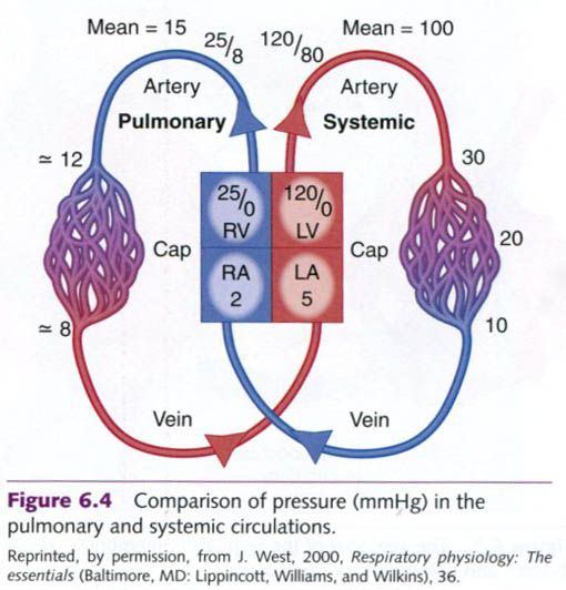
However, pressure and vascular resistance in the blood vessels in the lungs are
different than in the system circulation. The mean pressure in the pulmonary
artery is ~15 mmHg(systolic pressure is ~25 mmHg and diastolic pressure is ~8
mmHg) compared to the mean pressure in the aorta of ~95 mmHg. The pressure in
the left atrium where blood is returning to the heart from the lungs is ~5
mmHg; thus there is not a great pressure difference across the pulmonary
circulation(15-5 mmHg). Figure below illustrates the differences in pressures
between the pulmonary and systemic circulation.
Recalling the discussion of blood flow in the
cardiovascular system, pressure = flow x
resistance. Since blood flow to the lungs is equal to that of the systemic
circulation, and there is a substantially lower change in pressure across the
pulmonary vascular system, resistance is proportionally lower compared to that
in the systemic circulation. This is reflected in differences in the anatomy of
the vessels in the pulmonary versus systemic circulation: the pulmonary blood
vessels are thin walled, with relatively little smooth muscle.
Respiratory membrane
Gas
exchange between the air in the alveoli and the blood in the pulmonary
capillaries occurs across the respiratory membrane(also called the alveolar-capillary
membrane). This membrane,
depicted in the picture below, is composed of:
- The alveolar wall
- The capillary wall
- Their basement membranes.
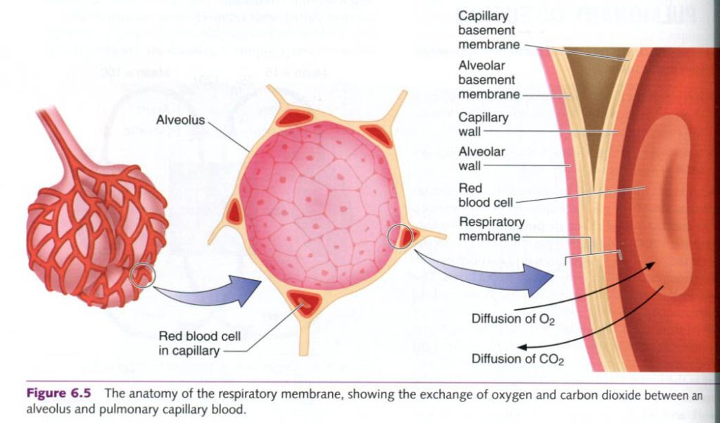
The primary function of these membranous
surfaces is for gas exchange. The respiration membrane is very thin, measuring
only 0.5 to 4.0 micrometres. As a result, the gases in the nearly 300 million
alveoli are in close proximity to the blood circulating through the
capillaries.
Partial pressure of gases
The air we breathe is a mixture of gases. Each
exerts a pressure in proportion to its concentration in the gas mixture. The
individual pressures from each gas in a mixture are reffered to as partial
pressures. According to Dalton
Consider the air we breathe. It is composed of
79.04% nitrogen(N2), 20.93% oxygen(O2), and 0.03% carbon
dioxide(CO2). These percentages remain constant regardless of
altitude. At sea level, the atmospheric(or barometric) pressure is
approximately 760 mmHg, which is also referred to as standard atmospheric
pressure. Thus, if the total atmospheric pressure is 760 mmHg, then the partial
pressure of nitrogen(PN2) in air is 600.7 mmHg(79.04% of the total
760 mmHg pressure). Oxygen’s partial pressure(PO2) is 159.1
mmHg(20.93% of 760 mmHg), and carbon dioxide’s partial pressure(PCO2)
is 0.2 mmHg(0.03% of 760 mmHg).
In the human body, gases are usually dissolved
in fluids, such as blood plasma. According to Henry’s law, gases
dissolve in liquids in proportion to their partial pressures, depending also on
their solubilities in the specific fluids and on the temperature. A gas’s
solubility in blood is a constant, and blood temperature also remains
relatively constant at rest. Thus, the most critical factor for gas
exchange between the alveoli and the blood is the pressure gradient between the
gases in the two areas.
Gas exchange in the alveoli
Differences in the partial pressures of the
gases in the alveoli and the gases in the blood create a pressure gradient
across the respiratory membrane. This forms the basis of gas exchange during
pulmonary diffusion. If the pressures on each side of the membrane were equal,
the gases would be at equilibrium and would not move. But the pressures are not
equal, so gases move according to partial pressure gradients.
Oxygen
exchange
The PO2 of air outside the body at
standard atmospheric pressure is 159 mmHg. But this pressure decreases to about
105 mmHg when air is inhaled and enters the alveoli, where it is moistened and
mixes with the air in the alveoli. The alveolar air is saturated with water
vapor(which has its own partial pressure) and contains more carbon dioxide than
the inspired air. Both the increased water vapor pressure and increased partial
pressure of carbon dioxide contribute to the total pressure in the alveoli.
Fresh air that ventilates the lungs is constantly mixed with the air in the
alveoli while some of the alveolar gases are exhaled to the environment. As a
result, alveolar gas concentrations remain relatively stable.
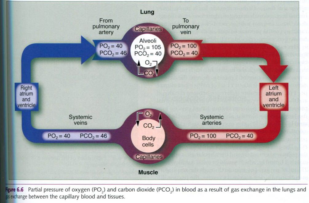
The blood, stripped of much of its oxygen by
the tissues, typically enters the pulmonary capillaries with a PO2
of about 40 mmHg(see figure below). This is about 60 to 65 mmHg less than the PO2
in the alveoli. In other words, the pressure gradient for oxygen across the
respiratory membrane is typically under 65 mmHg. As noted earlier, this
pressure gradient drives the oxygen from the alveoli into the blood to
equilibrate the pressure of the oxygen on each side of the membrane.
The PO2 in the alveoli stays
relatively constant at about 105 mmHg. As the deoxygenated blood enters the
pulmonary artery, the PO2 in the blood is only about the 40 mmHg.
But as the blood moves along the pulmonary capillaries, gas exchange occurs. By
the time the pulmonary blood reaches the venous end of these capillaries, the
PO2 in the blood equals that in the alveoli(approximately 105 mmHg),
and the blood is now considered to be saturated with oxygen as its full carrying
capacity. The blood leaving the lungs through the pulmonary veins and
subsequently returning to the systemic(left) side of the heart has a rich
supply of oxygen to deliver to the tissues. Notice, however, that the PO2
in the pulmonary vein is 100 mmHg, not the 105 mmHg found in the alveolar air
and pulmonary capillaries. This difference is attributable to the fact that
about 2% of the blood is shunted from the aorta directly to the lung to meet
the oxygen needs of the lung itself. This blood has a lower PO2 and
reenters the pulmonary vein along with fully saturated blood returning to the
left atrium that has just completed gas exchange. This blood mixes and thus
decreases the PO2 of blood returning to the heart.
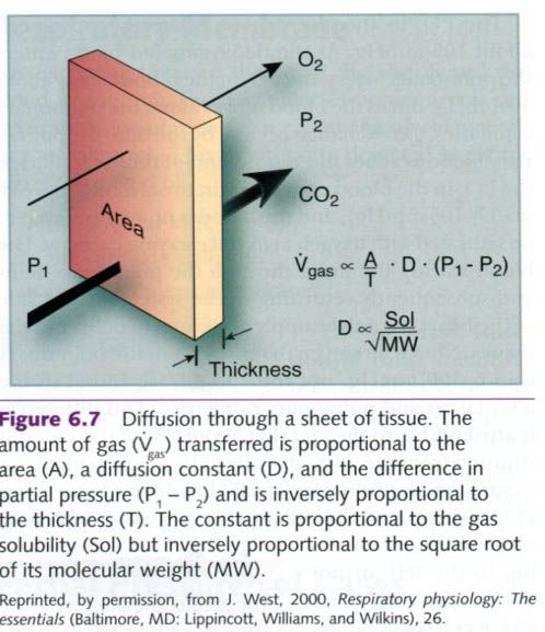
Diffusion through tissues is described by Fick’s law(see figure below). Fick’s law states that the rate of
diffusion through a tissue such as the respiratory membrane is proportional to
the surface area and the difference in the partial pressure of gas between the
two sides of the tissue. The rate of diffusion is also inversely
proportional to the thickness of the tissue in which the gas must diffuse.
Additionally, the diffusion constant, which is unique to each gas, influences
the rate of diffusion across the tissue. Carbon dioxide has a much lower
diffusion constant than oxygen; therefore, even though there is not as great a
difference between alveolar and capillary partial pressure of carbon dioxide as
there is for oxygen, carbon dioxide still diffuses easily.
The rate at which oxygen diffuses from the
alveoli into the blood is reffered to as the oxygen diffusion capacity and is
expressed as the volume of oxygen that diffuses through the membrane each
minute for a pressure difference of 1 mmHg. At rest, the oxygen diffusion
capacity is about 21ml of oxygen per minute per 1 mmHg of pressure difference
between the alveoli and the pulmonary capillary blood. Although the partial
pressure gradient between venous blood coming into the lung and the alveolar
air is about 65 mmHg(105 mmHg – 40 mmHg), the oxygen diffusion capacity is
calculated on the basis of the mean pressure in the pulmonary capillary, which
has a substantially higher PO2. The gradient between the mean
partial pressure of the pulmonary capillary and the alveolar air is
approximately 11 mmHg, which would provide a diffusion of 231ml of oxygen per
minute through the respiratory membrane. During maximal exercise, the oxygen
difference fusion capacity may increase by up to three times the resting rate,
because blood is returning to the lungs severely desaturated and thus there is
a greater partial pressure gradient from the alveoli to the blood. In fact,
rates of more than 80 ml/min have been observed among highly trained athletes.
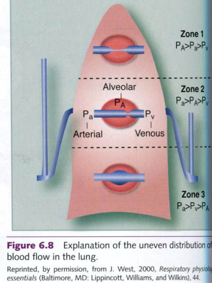
The increase in oxygen diffusion capacity from
rest to exercise is caused by a relatively inefficient, sluggish circulation
through the lungs at rest, which results primarily from limited perfusion of
the upper regions of the lungs attributable to gravity. If the lung is divided
into three zones as depicted in the figure below, at rest only the bottom
third(zone 3) of the lung is perfused with blood. During exercise, however,
blood flow through the lungs is greater, primarily as a result of elevated
blood pressure, which increases lung perfusion.
Carbon dioxide exchange
Carbon dioxide, like oxygen, moves along a
pressure gradient. As shown in the figure 3 of this draw, the blood passing
from the right side of the heart through the alveoli has a PCO2 of
about 46 mmHg. Air in the alveoli has a PCO2 of about 40 mmHg.
Although this results in a relatively small pressure gradient of only about 6
mmHg, it is more than adequate to allow for exchange of CO2. Carbon
dioxide’s diffusion coefficient is 20 times greater than that of oxygen, so CO2
can diffuse across the respiratory membrane much more rapidly.
The partial pressures of gases involved in
pulmonary diffusion are summarized in the table below. Note that the total
pressure in the venous blood is only 706 mmHg, 54 mmHg lower than the total
pressure in dry air and alveolar air. This is the result of a much greater
decrease in PO2 compared with the increase in PCO2 as the
blood goes through the body’s tissues.
|
Partial
pressure of gases at sea level
|
||||||
|
|
Partial
pressure (mmHg)
|
|||||
|
Gas
|
%
in dry air
|
dry
air
|
alveolar
air
|
arterial
blood
|
venous
blood
|
diffusion
gradient
|
|
H2O
|
0.00
|
0
|
47
|
47
|
47
|
0
|
|
O2
|
20.93
|
159.1
|
105
|
100
|
40
|
60
|
|
CO2
|
0.03
|
0.2
|
40
|
40
|
46
|
6
|
|
N2
|
79.04
|
600.7
|
568
|
573
|
573
|
0
|
|
Total
|
100.00
|
760
|
760
|
760
|
706
|
0
|


0 коментара:
Постави коментар