


- The P wave;
- The QRS complex;
- The T wave.
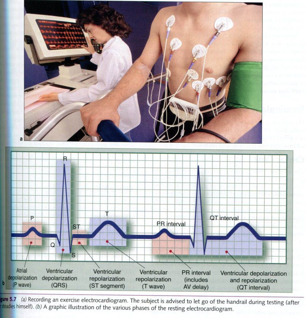
The P wave
represents atrial depolarization and occurs when the electrical impulse travels
from the SA node through the atria to
the AV node. The QRS complex represents ventricular depolarization and occurs as the
impulse spreads from the AV bundle to the Purkinje fibers and through the
ventricles. The T wave represents
ventricular repolarization. Atrial repolarization cannot be seen, because it
occurs during ventricular depolarization(QRS complex).
Electrocardiograms are often obtained during
exercise as clinical diagnostic tests of cardiac function. As exercise
intensity increases, the heart must beat faster and work harder to deliver more
blood to active muscles. Indications of coronary artery disease, not evident at
rest, may show up on the ECG as the heart increases its rate of work. Exercise
ECGs are also invaluable tools for research in exercise physiology because they
provide a convenient method for tracking heart rate and rhythm changes during
acute exercise.


0 коментара:
Постави коментар