


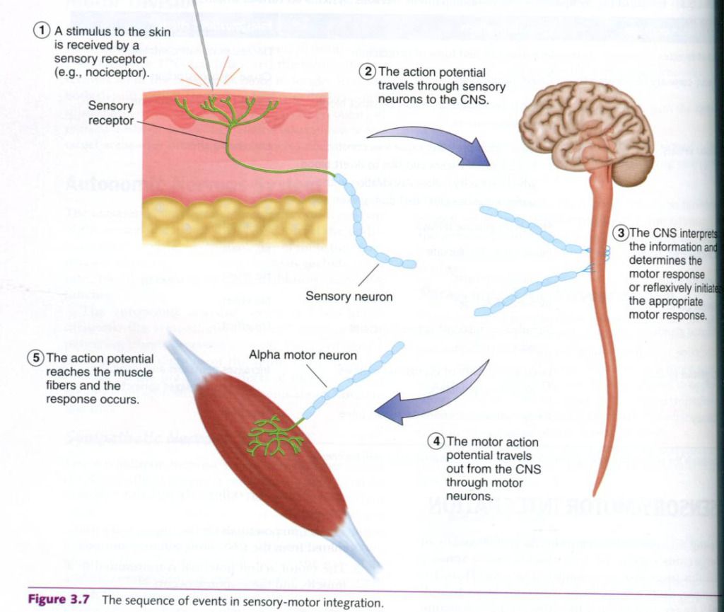
This process is called sensory-motor integration, and it is depicted in the figure below. For
the body to respond to sensory stimuli, the sensory and motor divisions of the
nervous system must function together in the following sequence of events:
- A sensory stimulus is
received by sensory receptors(e.g. pinprick).
- The sensory action potential
is transmitted along sensory neurons to the CNS.
- The CNS interprets the
incoming sensory information and determines which response is most
appropriate, or reflexively initiates a motor response.
- The action potentials for
the response are transmitted from the CNS along alpha-motor neurons.
- The motor action potential
is transmitted to a muscle, and the response occurs.
Sensory input
Recall that sensations and physiological status
are detected by sensory receptors throughout the body. The action potentials
resulting from sensory stimulation are transmitted via the sensory nerves to
the spinal cord. When they reach the spinal cord, they can trigger a local
reflex at that level, or they can travel to the upper regions of the spinal
cord or to the brain. Sensory pathways to the brain can terminate in sensory
areas of the brain stem, the cerebellum, the thalamus, or the cerebral cortex.
An area in which the sensory impulses terminate is referred to as in integration
center. This is where the sensory input is interpreted and linked to the motor
system. Figure below illustrates various sensory receptors and their nerve
pathways back to the spinal cord and up into various areas of the brain. The integration centers vary in function:
- Sensory impulses that terminate in the spinal cord are integrated
there. The response is typically a simple motor reflex, which is the
simpliest type of integration.
- Sensory signals that terminate in the lower brain stem result in
subconscious motor reactions of a higher and more complex nature than
simple spinal cord reflexes. Postural control during sitting, standing, or
moving is an example of this level of sensory input.
- Sensory signals that terminate in the cerebellum also result in subconscious
control of movement. The cerebellum appears to be the center of
coordination, smoothing out movements by coordinating the actions of the
various contracting muscle groups to perform the desired movement. Both
fine and gross motor movements appear to be coordinated by the cerebellum
in the concert with the basal ganglia. Without the control exerted by the
cerebellum, all movement would be uncontrolled and uncoordinated.
- Sensory signals that terminate at the thalamus begin to enter the
level of consciousness, and the person begins to distinguish various
sensations.
- Only when sensory signals enter the cerebral cortex can one
discretely localize the signal. The primary sensory cortex, located in the
postcentral gyrus(in the parietal lobe), receives general sensory input
from the receptors in the skin and from proprioceptors in the muscles,
tendons, and joints. This area has a map of the body. Stimulation in a
specific area of the body is recognized, and its exact location is known
instantly. Thus, this part of the conscious brain allows us to be
constantly aware of our surroundings and our relationship to them.

Motor control
Once a sensory impulse
is received, it may evoke a motor response, regardless of the level at which
the sensory impulse stops. This response can originate from any of three
levels:
·
The spinal cord
·
The lower regions of the brain
·
The motor area of the cerebral cortex
As of the level of control moves from the
spinal cord to the motor cortex, the degree of movement complexity increases
from simple reflex control to complicated movements requiring basic thought
processes. Motor responses for more complex movement patterns typically
originate in the motor cortex of the brain.
At last, we are ready to tie the two systems
together through sensory-motor integration. The simpliest form of this is the
reflex, so we can consider it first.
Reflex activity
What happens when one unknowingly puts one’s
hand on a hot stove? First, the stimuli of heat and pain are received by the
thermoreceptors and nocireceptors in the hand, and then sensory action
potentials travel to the spinal cord, terminating at the level of entry. Once
in the spinal cord, these action potentials are integrated instantly by
interneurons that connect the sensory and motor neurons. The action potentials
move to the motor neurons and travel to the effectors, the muscles controlling
the withdrawal of the hand. The result is that the person reflexively withdraws
the hand from the hot stove without giving the action any thought.
A motor
reflex is a preprogrammed response; any time the sensory nerves transmit
certain action potentials, the body responds instantly and identically. In
examples like the one just used, whether one touches something that is too hot
or too cold, thermoreceptors will elicit a reflex to withdraw the hand. Whether
the pain arises from heat or from a sharp object, the nociceptors will also
cause a withdrawal reflex. By the time one is consciously aware of the specific
stimulus(after sensory action potentials also have been transmitted to the
primary sensory cortex), the reflex activity is well under way, if not
completed. All neural activity occurs extremely rapidly, but a reflex is the
fastest mode of response because the impulse is not transmitted up the spinal
cord to the brain before an action occurs. Only one response is possible; no
options need to be considered.
Muscle spindles
Now that we have covered the basics of reflex
activity, we can look more closely at two reflexes that help control muscle
function. The first involves a special structure: the muscle spindle.
The muscle
spindle lies between regular skeletal muscle fibers, referred to as extrafusal(outside the spindle) fibers.
A muscle spindle consists of 4 to 20 small, specialized muscle fibers called intrafusal(inside the spindle) fibers
and the nerve endings, sensory and motor, associated with these fibers. A
connective tissue sheath surrounds the muscle spindle and attaches to the
endomysium of the extrafusal fibers. The intrafusal fibers are controlled by
specialized motor neurons, reffered to as gamma-motor
neurons. In contrast, extrafusal fibers(the regular fibers) are controlled
by alpha-motor neurons.
The central region of an intrafusal fiber
cannot contract because it contains no or only a few actin and myosin
filaments. Thus, the central region can only stretch. Because the muscle
spindle is attached to the extrafusal fibers, any time those fibers are
stretched, the central region of the muscle spindle is also stretched.
Sensory nerve endings wrapped around this
central region of the muscle spindle transmit information to the spinal cord
when this region is stretched, informing the CNS of the muscle’s length. In the
spinal cord, the sensory neuron synapses with an alpha-motor neuron, which
triggers reflexive muscle contraction(in the extrafusal fibers) to resist
further stretching.
Let’s illustrate this action with an example. A
person’s arm is bent at the elbow, and the hand is extended, palm up. Suddenly
someone places a heavy weight in the palm. The forearm starts to drop, which
stretches the muscle fibers in the elbow flexors, which in turn stretch the
muscle spindles. In response to that stretch, the sensory neurons send action
potentials to the spinal cord, which then activates the alpha-motor neurons of
motor units in the same muscles. These cause the muscles to increase their
force production overcoming the stretch.
Gamma-motor neurons excite the intrafusal
fibers, prestretching them slightly. Although the midsection of the intrafusal
fibers cannot contract, the ends can. The gamma-motor neurons cause slight
contraction of the ends of these fibers, which stretches the central region
slightly. This prestretch makes the muscle spindle highly sensitive to even
small degrees of stretch.
The muscle spindle also assists normal muscle
action. It appears that when the alpha-motor neurons are stimulated to contract
the extrafusal muscle fibers, the gamma-motor neurons are also activated,
contracting the ends of the intrafusal fibers. This stretches the central
region of the muscle spindle, giving rise to sensory impulses that travel to
the spinal cord and then to the alpha-motor neurons. In response, the muscle
increases its form production. Thus, muscle force production is enhanced through
this function of the muscle spindles.
Information brought into the spinal cord from
the sensory neurons associated with muscle spindles does not merely end at that
level. Impluses are also sent up to higher parts of the CNS, supplying the
brain with continuous feedback on the exact length of the muscle and the rate
at which that length is changing. The information is essential for maintaining
muscle tone and posture and for executing movements. The muscle spindle
functions as a servo-mechanism to continuously correct movements that do no
proceed as planned. The brain is informed of errors in the intended movement at
the same time that the error is being corrected at the spinal cord level.
Golgi Tendon organs
Golgi
tendon organs are encapsulated
sensory receptors through which a small bundle of muscle tendon fibers pass.
These organs are located just proximal to the tendon fibers’ attachment to the
muscle fibers, as shown in the figure below. Approximately 5 to 25 muscle
fibers are usually connected with each Golgi tendon organ. Whereas muscle
spindles monitor the length of a muscle. Golgi tendon organs are sensitive to
tension in the muscle-tendon complex and operate like a strain gauge, a device
that senses changes in tension. Their sensitivity is so great that they can
respond to the contraction of a single muscle fiber. These sensory receptors
are inhibitory in nature, performing a protective function by reducing the
potential for injury. When stimulated, these receptors inhibit the
contracting(agonist) muscles and excite the antagonist muscles.
Some researchers speculate that reducing the
influence of Golgi tendon organs disinhibits the active muscles, allowing a
more forceful muscle action. This mechanism may explain at least part of the
gains in muscular strength that accompany strength training.


0 коментара:
Постави коментар