


Arteries are large muscular, elastic, conduit vessels
for transporting blood away from the heart to the arterioles. The aorta is the major artery transporting
blood from the left ventricle to all regions of the body, and it branches into
smaller arteries that become progressively smaller, finally branching into
arterioles. The arterioles are the site of greatest control of the circulation
by the sympathetic nervous system, so arterioles are sometimes called
resistance vessels.
From
the arterioles, blood enters the capillaries, the narrowest vessels, with walls
only one cell thick. Virtually all exchange between the blood and the tissues
occurs at the capillaries. Blood leaves the capillaries to begin the return
trip to the heart in venules, and the venules form larger vessels – the veins.
The vena cava is the great vein transporting blood back to the right atrium
from all regions to the body above(superior vena cava) and below(inferior vena
cava) the heart.
Blood pressure
Blood pressure is the pressure exerted by the
blood on the vessel walls, and the term usually refers to arterial blood
pressure. It is expressed by two numbers: the systolic blood pressure(SBP) and the diastolic blood pressure(DBP).
The higher number is the SBP; it represents the highest pressure in the artery
that occurs during ventricular systole. Ventricular contraction pushes the
blood through the arteries with tremendous force, and that force exerts high
pressure on the arterial walls. The lower number is the DBP and represents the
lowest pressure in the artery, corresponding to ventricular diastole when the
ventricle is filling.
Mean
arterial pressure(MAP) represents
thee average pressure exerted by the blood as it travels through the arteries.
Since diastole takes about twice as long as systole in a normal cardiac cycle,
mean arterial pressure can be estimated from the DBP and SBP as follows:
MAP = 2/3 DBP + 1/3 SBP
Alternately,
MAP = DBP + [0.333 x (SBP – DBP)].
(SBP – DBP) is also called pulse pressure.
To illustrate, with a normal resting blood
pressure of 120 mmHg over 80 mmHg, the MAP = 80 + [0.333 + (120-80)] = 93 mmHg.
General hemodynamics
Recall that the cardiovascular system is a
continuous closed-loop system. Blood flows in this closed-loop system because
of the pressure gradient that exists between the arterial and venous sides of
the circulation. To understand regulation of blood flow to the tissues it is
necessary to understand the intricate relationship between pressure, flow and
resistance.
In order for blood to flow in a vessel there
must be a pressure difference from one end of the vessel to the other end.
Blood will flow from the region of the vessel with high pressure to the region
of the vessel with low pressure. Alternatively, if there is no pressure
difference across the vessel, there is no driving force and therefore no blood flow.
In the circulatory system, the mean arterial pressure in the aorta is
approximately 100 mmHg at rest, and the pressure in the right atrium is very
close to 0 mmHg. Therefore, the pressure difference across the entire
circulatory system is 100 mmHg – 0 mmHg = 100 mmHg.
The
reason for the pressure differential from the arterial to the venous
circulation is that the blood vessels themselves provide resistance or
impedance to blood flow. The resistance that the vessel provides is largely
dictated by the properties of the blood vessels and the blood itself. These
properties include the length and radius of the blood vessel and the viscosity
of thickness of the blood flowing through the vessel. Resistance to flow can be
calculated as
resistance
= [ἠL/r4]
where
ἠ is the viscosity of the blood, L is the length of the vessel, and r is the
radius of the vessel, which is raised to the fourth power.
Blood
flow is proportional to the pressure difference across the system and is
inversely proportional to resistance. This relationship can be illustrated by
the following equation:
blood
flow = ▲pressure/ resistance
Notice
that blood flow can increase by either an increase in the pressure
difference(▲pressure), a decrease in resistance, or a combination of the two. Altering
resistance to control blood flow is much more advantageous because very small
changes in blood vessel radius equate to large changes in resistance. This is
due to the fourth-power mathematical relationship between vascular resistance
and vessel radius.
Changes in vascular resistance are largely due
to changes in blood vessel radius or diameter, as the viscosity of blood and
the length of the vessels do not change significantly under normal conditions.
Therefore, regulation of blood flow to organs is accomplished by small changes
in blood vessel radius through vasoconstriction
and vasodilatation. This allows the cardiovascular system to divert blood
flow to the areas where it is needed most.
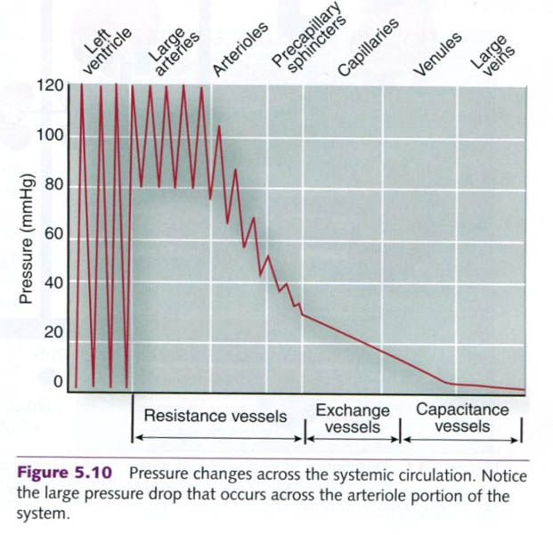
As mentioned already, most resistance to the
blood flow occurs in the arterioles. Figure below shows the blood pressure
changes across the entire vasculary system. The arterioles are responsible for
-70% to 80% of the drop in mean arterial pressure across the entire
cardiovascular system. This is important because small changes in arteriole
radius can greatly affect the regulation of mean arteriole pressure and the
local control of blood flow. At the capillary level, changes due to systole and
diastole are no longer evident and the flow is smooth(laminar) rather than
turbulent.
Distribution of blood
Distribution of blood to the various body
tissues varies tremendously depending on the immediate needs of a specific
tissue compared with that of other areas of the body. At rest under normal
conditions, the most metabolically active tissues receive the greatest blood
supply. The liver and kidneys combine to receive almost half the blood being
circulated, and resting skeletal muscles receive only about 15% to 20%.
During exercise, blood is redirected to the
areas where it is needed most. During heavy endurance exercise, muscles receive
up to 80% or more of the available blood. This redistribution, along with
increases in cardiac output, allows
up to 25 times more blood flow to active muscles.

Similarly, after one eats a big meal, the
digestive system receives more of the available cardiac output than when the
digestive system is empty. Along the same lines, during increasing
environmental heat stress, skin blood flow increases to a greater extent as the
body attempts to maintain normal temperature. The cardiovascular system
responds accordingly to redistribute blood, whether it is to the exercising
muscle to match metabolism, for digestion, or to facilitate thermoregulation.
These changes in the distribution of the cardiac output are controlled by the
sympathetic nervous system, primarily by increasing or decreasing arteriolar
diameter. These vessels have a strong muscular wall that can significantly
after vessel diameter, are highly innervated by sympathetic nerves, and have
the capacity to respond to local control mechanisms.
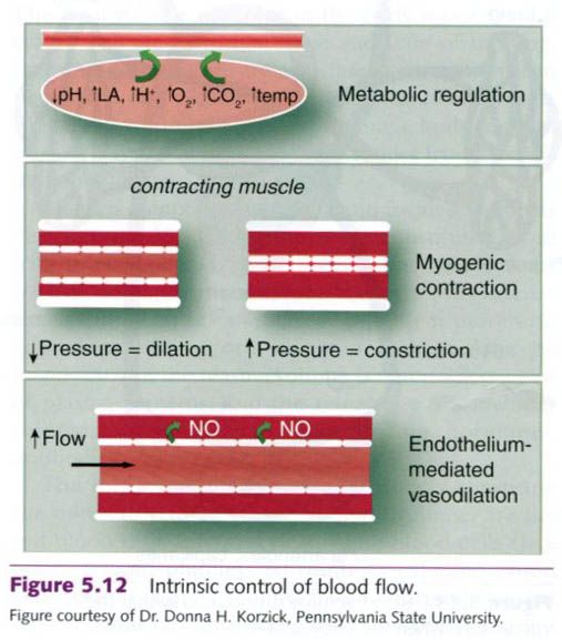
Intrinsic control of blood flow
Intrinsic
control of blood distribution refers to the ability of the local tissues to
vasodilate or vasoconstrict the arterioles that serve them and alter regional
blood flow depending on the immediate needs of those tissues. With exercise and
the increased metabolic demand of the exercising skeletal muscles, the
arterioles undergo locally mediated vasodilation, opening up to allow more
blood to enter that highly active tissue.
There are essentially three types of intrinsic
control of blood flow. The strongest stimulus for the release of local
vasodilating chemicals is metabolicm in particular an increased oxygen demand.
As the tissue’s oxygen use increases, available oxygen is diminished. Local
arterioles vasodilate to allow more blood, and thus more oxygen, to perfuse
that area. Other chemical changes that can stimulate increased blood flow are
decreases in other nutrients and increases in by-products(carbon dioxide, K+,
H+, lactic acid) or inflammatory chemicals. Second, several
vasodilating substances can be produced in the endothelium(inner lining) of
arterioles and initiate vasodilation in the vascular smooth muscle of the
arterioles. These substances include nitric oxide(NO), prostaglandins, and
endothelium-derived hyperpolarizing factor(EDHF). These endothelium-derived
vasodilators are important to the regulation of blood flow at rest and during
exercise in humans, although the precise mechanisms and interplay between these
vasodilators are still being studied. Finally, pressure changes within the
vessels themselves can also cause vasodilation and vasoconstriction. This is
reffered as the myogenic response. The vascular smooth muscle contracts in
response to an increase in pressure across the vessel wall and relaxes in
response to a decrease in pressure across the vessel wall. Additionally,
acetylcholine and adenosine also have been proposed as potential vasodilators
for the increase in muscle blood flow during exercise. Increased blood flow can
either bring in needed substances such as oxygen, or clear out metabolic waste
such as carbon dioxide, or usually both. Figure below illustrates the three
types of intrinsic control of vascular tone.
Extrinsic neural control
This
concept of intrinsic local control explains redistribution of blood within an
organ or tissue mass; however, the cardiovascular system must divert blood flow
to where it is needed, beginning at a site upstream of the local environment.
Redistribution at the system or body level is controlled by neural mechanisms.
This is known as extrinsic neural control of blood flow, because the control
comes from outside the specific area(extrinsic) instead of from locally inside
the tissues(intrinsic).
Blood flow to all body parts is regulated
largely by sympathetic nervous system. The circular layers of smooth muscle
within the artery and arteriole walls are supplied by sympathetic nerves. In
most vessels, an increase in sympathetic nerve activity causes these muscle
cells to contract, constricting blood vessels and thereby decreasing blood
flow.
Under normal conditions, the sympathetic nerves
transmit impulses continuously to the blood vessels, keeping the vessels
moderately constricted to maintain adequate blood pressure. This state of tonic
vasoconstriction is referred to as vasomotor tone. When sympathetic stimulation
increases, further constriction of the blood vessels in a specific area
decreases blood flow into that area and allows more blood to be distributed
elsewhere. But if sympathetic stimulation decreases below the level needed to
maintain tone, constriction of vessels in the area is lessened, so the vessels
passively vasodilate, increasing blood flow into that area. Therefore, sympathetic
stimulation will cause vasoconstriction in most vessels, but blood flow is
altered by either increasing or decreasing the amount of vasoconstriction
relative to normal vasomotor tone.
Distribution of venous blood
While
flow to tissues is controlled by changes on the arterial side of the system,
most of the blood volume normally resides in the venous side of the system. At
rest, the blood volume is distributed among the vascular shown in the figure
below. The venous system has a great capacity to hold blood volume. There is a
little vascular smooth muscle in the veins, and they are very elastic and
“balloon-like”. Thus, the venous system provides a large reservoir of blood
available to be rapidly distributed back to the heart(venous return) and to the
arterial circulation. This is accomplished through sympathetic stimulation of
the venules and veins, which causes the vessels to constrict.

Integrative control of blood pressure
Blood pressure is normally maintained by
reflexes from the autonomic nervous system. Specialized pressure sensors
located in the aortic arch and the carotid arteries, called baroreceptors, are sensitive to changes
in arterial pressure. They send information about the current blood pressure to
the cardiovascular control centers in the brain where autonomic reflexes are
initiated to respond to changes in blood pressure. For example, when blood
pressure is elevated, the baroreceptors are stimulated by an increase in
stretch. They relay this information to the cardiovascular control center in
the brain. In response to the increased pressure there is a reflex increase in
vagal tone, to decrease heart rate, and a decrease in sympathetic activity,
which serves to normalize blood pressure. In response to a decrease in blood
pressure, less stretch is sensed by the baroreceptors, and the response is to
increase heart rate by vagal withdrawal and to increase sympathetic nervous
activity, thus correcting the low-pressure signal.
There are also other specialized receptors,
called chemoreceptors and mechanoreceptors, that send information
about the chemical environment in the muscle and the length and tension of the
muscle to the cardiovascular control centers. These receptors can also modify
the blood pressure response and are especially important during exercise.
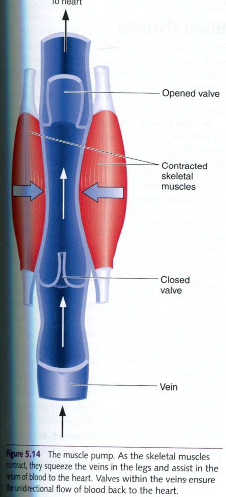
Return of blood to the heart
Because we spend so much time in an upright
position, the cardiovascular system requires mechanical assistance to overcome
the force of gravity when blood returns from the lower parts of the body to the
heart. Three basic mechanisms assist in this process:
- Valves in the veins
- The muscle pump
- The respiratory pump.
The veins contain valves that allow blood to
flow in only one direction, thus preventing backflow and pooling of blood in
the lower body. These venous valves also complement the action of the skeletal
muscle pump, mechanical compression of the veins from rhythmic skeletal muscle
contraction. This pushes blood volume in the veins back toward the heart.
Finally, the changes in pressure in the abdominal and thoracic cavities during
breathing assist blood return to the heart.


0 коментара:
Постави коментар