


Gluteus
maximus
|
Semitendinosus
|
HAMSTRINGS
|
|
Semimbranosus
|
|
|
Biceps
femoris
|
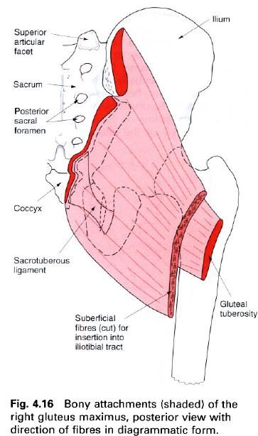
Gluteus maximus
As its name implies, this is the largest of the
gluteal muscles. It is very powerful and is situated on the posterior aspect of
the hip joint, being responsible for
the very pleasant shape of this region in humans. In lower primates gluteus
maximus in an adductor of the hip;
this was also the case in early, primitive humans. However, with the changes
that have occurred in the human pelvis to enable the erect posture to be
adopted, the muscle has become mainly an extensor of the hip. It is the muscle mainly responsible for the erect position,
thereby freeing the forelimbs from a weight-bearing role, and enabling them to
become the precision implements that they are today.
Gluteus maximus is quadrilateral in shape
consisting of bundles of muscle fibers laid down in the line of pull of the
muscle, giving the surface of the muscle a coarse appearance. The muscle is
thick and forms two layers as it passes down to its lower attachment. Above, it
attaches to the gluteal surface of
the ilium behind the posterior
gluteal line, the posterior border of
the ilium and from the adjacent part
of the iliac crest. It also arises
from the side of the coccyx and the posterior aspect of the sacrum, including the upper part of the sacrotuberous ligament. Its upper fibres
attach to the aponeurosis of the sacrospinalis while its deep anterior
fibres come from the fascia which covers gluteus medius. Although this appears
to be a vast area, it must be remembered that most of these structures can be
covered with one hand if it were
slipped into the back pocket of a pair of trousers.
The fibres pass downwards and forwards towards
the upper end of the femur. The most
superficial, about three-quarters, of the fibres form a separate lamina, which
narrows down and attaches between the two layers of the fascia lata helping to
form the iliotibial tract. The deeper
remaining one-quarter of the muscle fibres form a broad aponeurosis which
attaches to the gluteal tuberosity of
the femur.
Nerve
supply
Gluteus maximus is supplied by the inferior gluteal nerve, root value L5,
S1, 2. The skin covering the muscle, however, is mainly supplied by branches
from L2 and S3.
Action
When acting from above, the muscle pulls the
shaft of the femur backwards
producing extension of the flexed hip joint. As its lower attachment is nearer
to the lateral side of the thigh, the muscle will tend to rotate the thigh
laterally during extension. The lower fibres can adduct the thigh, while the
upper fibres may help in abduction.
The fibres which attach to the iliotibial tract
can produce extension of the knee joint because the lower end of the tract
attaches to the lateral tibial condyle anterior to the axis of movement.
Through the iliotibial tract, gluteus maximus provides powerful support on the
lateral side of the knee. If the femur
is fixed, contraction of gluteus maximus will pull the ilium and pelvis backwards around the hip joint, but this time the pelvis and trunk are the moving parts,
and a lifting of the trunk from a flexed position occurs.
Functional
activity
Being a powerful extensor of the thigh,
especially when the hip has been flexed, means that this muscle is ideally
suited for fulfilling its role in such powerful movements as stepping up onto a
stool, climbing and running. However, it is not used greatly as an extensor in
ordinary walking.
With the hamstrings, it will take part in
raising the trunk from a flexed position, as in standing upright from a bent
forward position. Indeed, gluteus maximus and the hamstrings provide the main
control in forward bending of the body, as the movement primarily occurs at the
hip joint.
It plays an important role in balancing the pelvis
on the femoral heads thus helping to maintain the upright posture; its ability
to aid lateral rotation of the femur
when standing assists in raising the medial longitudinal arch of the foot.
The role of gluteus maximus during sitting
should not be dismissed. Although the ischial tuberosities support the majority
of the weight of the trunk when sitting, pressure is regularly relieved from
these bony points by a static or sometimes dynamic contraction of the muscle
which raises the tuberosities of the ischium from the supporting surfaces. The
muscle is then relaxed and the weight is then lowered. Sometimes the weight is
shifted from side to side with the alternate use of gluteus maximus of each
side.
Paralysis of the muscle will lead to a
flattening of the buttock with a loss of the beautiful contour, and an
inability to climb stairs and run. However, it must be kept in mind that there
are other muscles which can be brought into action to produce extension of the hip, although it is much weaker
movement. Gluteus maximus can be developed to produce a functional extension of
the knee in patients where quadriceps femoris is either very weak or paralysed.
This is not a powerful movement, but may be sufficient to enable the patient to
extend the knee and enable the lower limb to become weight-bearing during
walking or standing.
Palpation
On a model or on yourself, first find some bony
prominences which will give useful landmarks of the muscle. The iliac crest is
easily palpated approximately at the belt level; moving the hand backwards along the crest a small
bony process can be felt: this is the posterior superior iliac spine. With the
fingers running inferiorly and medially let this be the centre of the palm. The
hand will now just about cover the
upper attachment of gluteus maximus; the palm is over the posterior part of the
ilium, the sacrum and the back of the sacroiliac joint, while the tips of
the fingers are on the edge of the coccyx
and the upper end of the sacrotuberous ligament. The bulk of the muscle is now
under the palm, follow this path to the greater trochanter of the femur. Now try the following:
- Extend the lower limb whilst in the standing position, keeping the hand on the muscle; it goes hard and produces a much clearer shape.
- Place the foot onto a stool and put the hand in the same position as before and step up. Again the muscle will be felt coming into action very strongly.
- Take up the standing position and place the hands on each gluteus maximus as if they were in the back pocket. Raise the medial borders of the feet as if to shorten the medial longitudinal arch of the foot. As the arch is raised gluteus maximus will be felt working quite strongly, with the femur tending to rotate laterally.
- Finally, take up the sitting position but this time place a hand under each buttock so that the ischial tuberosity now rests on the hand. Now move the weight from side to side as if getting tired of sitting. Gluteus maximus now contacts alternately, taking the weight off the tuberosity and then lowering it down again.
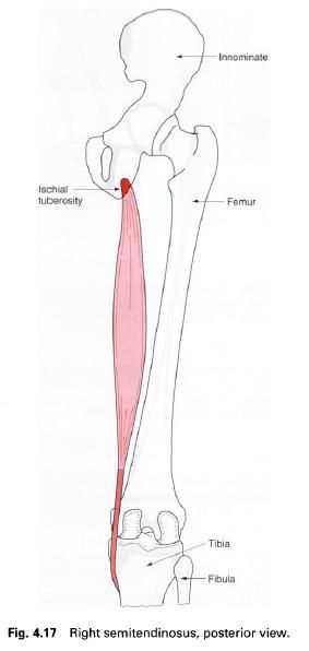
Semitendinosus
The upper attachment of this muscle is from the
lower medial facet of the lateral
section of the ischial tuberosity.
Its tendon of attachment is combined with that of the biceps femoris and the
two muscles run together for a short distance. It then forms a fusiform muscle
belly which quickly gives way to a long tendon, thus accounting for its name.
This tendon passes downwards and medially behind the medial condyle of the femur, being separated from the medial
collateral ligament by a small bursa, to attach to a vertical line on the
medial surface of the medial condyle
of the tibia just behind the
insertion of the sartorius and behind and below the attachment of the gracilis.
Near its insertion it is separated from gracilis is separated from sartorius by
another bursa.
Nerve
supply. Semitendinosus is
supplied by the tibial division of the
sciatic nerve, root value L5, S1, 2. The skin covering the muscle is
supplied mainly by S2.
Action. Semitendinosus will, when working from below,
help to extend the hip joint when the trunk is bent forward. When working from
above it will aid in flexion of the knee joint; if the knee is semiflexed it
will produce medial rotation of the knee. If the foot is fixed, semitendinosus
will act as a lateral rotator of the femur
and pelvis on the tibia.
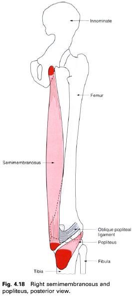
Semimembranosus
This muscle is situated on the posteromedial
side of the thigh in its lower part, deep to semitendinosus. It attaches by a
strong membranous tendon to the upper
lateral facet on the rough part of the ischial
tuberosity and passes downwards and medially. The muscle becomes fleshy on
the medial side of the tendon, being deep to semitendinosus and biceps femoris.
From the lower part of the muscle a second aponeurotic tendon arises narrowing
down towards its lower attachment, which is a horizontal groove on the posteromedial surface of the medial tibial
condyle. From here its fibres spread in all directions, but particularly
upwards and laterally forming the oblique popliteal ligament. Bursae separate
the muscle from the medial head of gastrocnemius and from the tibia near its attachment.
Nerve
supply. Semimembranosus is
supplied from the tibial division of the sciatic nerve, root value L5, S1, 2.
The nerve supply to the skin covering the muscle is the same as that for the
semitendinosus, that is mainly from S2.
Action. As for semitendinosus.
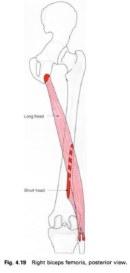
Biceps femoris
Biceps
femoris is situated on the posterolateral aspect of the thigh, arising by two
heads as the name implies, these being separated by a considerable distance.
The long head attaches to the lower medial facet on the ischial tuberosity with the tendon of
the semitendinosus, spreading on to the sacrotuberous
ligament. These two tendons descend together for a short distance then
separate into the two individual muscles, the long head of biceps forming a
fusiform muscle running downwards and laterally across the posterior aspect of
the thigh superficial to the sciatic nerve. In the lower third of the thigh the
long head begins to narrow and is joined on its deep aspect by the short head
of biceps.
The short head has its upper attachment from
the lower half of the lateral lip of the linea aspera reaching almost as far up as the attachment of gluteus
maximus and running down onto the upper
half of the lateral supracondylar
line of the femur; some fibres
arise from the lateral intermuscular septum. The fibres of this short head
gradually blend with the narrowing tendon of the long head which lies
superficial to it.
On approaching the knee, the tendon can be felt
crossing its posterolateral aspect running towards the head of the fibula.
Prior to its attachment to the head of the fibula the tendon of biceps
femoris is split in two by the fibular collateral ligament. Some fibres of the
tendon join the ligament, while a few others attach to the lateral tibial
condyle and some to the posterior aspect of the lateral intermuscular septum
which lies just in front of it. A bursa separates the tendon from the fibular
collateral ligament.
Nerve
supply. The long head of
biceps femoris is supplied by the tibial
portion of the sciatic nerve, while the short head is supplied by the common peroneal portion; the root value
of both is L5, S1, 2. The skin covering the muscle is supplied mainly by S2.
Action. Biceps femoris helps the previous three
muscles to extend the hip joint, particularly when the trunk is bent forwards
and is to be raised to the erect position. All three hamstrings will, of
course, control the lowering forward of the trunk; however in this case they
are working eccentrically, that is working, but its two ends moving apart.
Biceps femoris aids the semimembranosus and semitendinosus muscles in flexing
the knee joint. With the knee in a semiflexed position biceps femoris will
rotate the leg laterally on the thigh or if the foot is fixed, medially rotate the thigh and pelvis on the leg.
The hamstrings
Semitendinosus, semimbranosus and biceps femoris
are collectively known as the hamstrings.
They make up the large mass of muscle which can
be palpated on the posterior aspect of the thigh, and are involved with
extension of the hip, flexion of the
knee and rotation of the flexed knee. When working from above they can either
flex the knee as a group, or they can work individually in rotating the flexed
knee. If they are working with the lower attachment fixed, they will act as a
group extending the hip joint.
Functional
activity of the hamstrings
All three muscles cross the posterior aspect of
both the hip and the knee joints. Flexion of the knee and their stabilizing
effect is no doubt a very important function of these muscles, although for
this action a much smaller muscle bulk would have been sufficient. Extension of
the hip joint when the thigh is the moving part would also require a far
smaller group of muscles, especially when it is remembered that gluteus maximus
is far better situated to do the job. Raising the trunk from a flexed position
on the other hand requires a great deal more power as the muscles are working
with a very short lever arm: the ischium and its ramus. The weight of the trunk
acting on the other side of the hip
joint is considerable.
The mode of action of this group of muscles may
well be the reason it is injured so frequently during sports activities. The
most common cause of sports injury appears to be in the running section of
athletics, being more common in the first 10-20m of a sprint. This is often
blamed on inadequate preparation and warm-up before the start, and to some
extent this may be true. It must, however, be remembered that at this stage in a
race the hamstrings are contracting strongly and are acting over two joints.
At the start of a race the athlete is in a
forward-lean position in order to gain as much forward motion as possible.
Starting blocks serve to increase the degree of forward leaning. The hamstrings
are therefore working to their maximum, either to raise the trunk to an upright
position, or to hold the trunk in such a position that forward collapse of the
body as a whole is imminent. At the same time the lower limb is being thrust
forward to gain as much ground as possible, with flexion of the knee to prevent
the foot touching the ground. The
hamstrings must be under immense strain in this position and it is not
surprising that the muscle may tear.
The hamstrings play an important part in the
fine balancing of the pelvis in the standing position, particularly when the
upper trunk is being moved off the vertical axis. Working in conjunction with
the abdominal muscles anterosuperiorly and gluteus maximus posteroinferiorly,
the anteroposterior tilt of the pelvis can be altered. This will have an effect
on the lumbar lordosis.
Finally, the hamstrings have a role in decelerating the forward
motion of the tibia when the free swinging leg is extended during walking, and
so prevents the knee snapping into extension.


0 коментара:
Постави коментар