


The human lower limb is adapted for
weight-bearing, locomotion and maintaining the unique, upright, bipedal
posture. For all of these functions much more strength and stability are
required than in the upper limb. The bones of the lower limb are larger and
more robust than their upper limb counterparts, and vary in their
characteristics in relation to muscular development and body build. Many bones,
particularly the innominate and, to a lesser extent the femur, show sexual differences;
variations in the female pelvis for example being an adaptation to
child-bearing.
The form and structure of individual bones are
adapted to the function of support and resisting mechanical stresses. The
internal architecture of the bone is arranged to resist all such stresses and
forces. This is particularly marked at the articular regions of the bone.
During growth and life continuous modifications are being made in order to
maintain the functions of support and resistance to stress as the stresses
change. The attainment of an habitual, upright posture and bipedal gait has
resulted in both the mechanical and functional requirements of all the bones of
the lower limb. Consequently, during evolution, there have been major changes
in the lower limb.
The pelvic girdle, formed by the innominate of
each side articulating anteriorly at the symphysis pubis, connects the lower
limb to the vertebral column via its posterior articulation with the sacrum.
This posterior articulation, the sacroiliac joint, provides great strength in
the region of weight transference from the trunk to the lower limb at the
sacrifice of almost all mobility. The human ilium has developed so that it is
no longer blade-like but is shortened and tightly curved backwards and outwards,
changing the actions of the gluteal muscles. The changes in the pelvis have
resulted in a shift in its positioning from an essentially horizontal to an
essentially vertical position. This has enabled the trunk to be held
vertically, but has necessitated a change in the orientation of the sacrum with
respect to the ilium with the result that the axis of the pelvic canal lies
almost at a right angle to the vertebral column. During evolution there has
been a relative approximation of the sacral articular surface to the acetabulum
which makes for greater stability in the transmission of the weight of the
trunk to the hip joint. The increase in the magnitude of this weight has
resulted in an increase in the area of contact between sacrum and ilium
relative to the area of the ilium as a whole. For the same reason the
acetabulum and femoral head have also increased in relative size during
evolution. The shortening of the ischium that has occurred is an adaptation for
speed and rapid movements, which is of great importance in bipeds. Thus power
of action has been sacrified for speed.

Changes have also occurred at the knee with the
femoral condyles being more parallel in humans as compared with other primates.
The major change though has been in bringing the knees inwards towards the body
midline, which appears to be part of the overall pattern of centring the body
mass, thus reinforcing skeletal rather than muscular equilibrium.
In humans, the tibia and fibula are held
tightly together, with the tibia being the weight-bearing component while the
fibula is mainly for muscle attachments. There has been a loss of rotation of
the fibula with respect to the tibia.
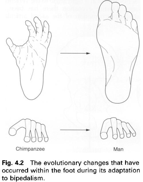
It is the foot, however, which has perhaps
undergone the greatest change during evolution. These changes reflect not so
much the evolution of a new function as a reduction in the original primate
functions, with the foot changing from a grasping, tactile organ to a locomotor
prop. Although some non-locomotor function is still possible, the foot has
evolved from a generalized to a specified organ. The joints of the human foot
permit much less internal mobility – an adaptation to ground walking. In
locomotion, the foot acts as a lever adding propulsive force to that of the
leg, with the point of pivot being the subtalar joint. The front part of the
foot has been shortened relative to the posterior portion, where the main
thrust in walking is developed – the power capabilities are thus accentuated.
The arches of the foot are formed by the shape
of the bony elements, and are supported by ligaments and tendons. They convert
the foot into a complex spring under tension and allow it to transmit the
stresses involved in walking, both when body momentum is checked at heel-strike
and when the foot is used in propelling the body forward. The lateral arch
helps to steady the foot on the ground, while the medial arch transmits the
main force of thrust in propulsion. This arched form of the foot is important
in providing one of the major determinants of gait, helping to minimize energy
expenditure and thus increase the efficiency of walking.
An important consequence of the upright,
bipedal posture is that the centre of gravity of the body has been brought
towards the vertebral column, so that in humans it lies slightly behind and at
about the same level as the hip joint thereby reducing the tendency of gravity
to pull the trunk forward. The centre of gravity projection then passes
anterior to the knee and ankle joints. At the knee, the line of weight
transmission passes towards the outside. Because of the angulation of the
femur, during walking of the foot, tibia and knee joint of each leg stay close
to the line followed by the centre of gravity, and thus energy expenditure is
minimal in maintaining the centre of gravity above the supporting limb. Balance
is thus improved and there is more time for the free leg to swing forward
enabling an increase in stride length. The alteration in the line of weight
transmission is carried into the foot, where it passes to the inner side. However,
it must be remembered that weight is also transmitted through the outside of
the foot, bringing the entire foot into use as a stabilizing element.
In order to reduce the possibility of collapse
or dislocation, due to the forces to which they are subjected, the joints of
the lower limb are structurally more stable than those of the upper limb. This
stability is either due to the shape of the articular surfaces, the number and
strength of the ligaments, or the size of the muscles related to the joint.
Commonly, each of these factors contributes to a varying extent.


0 коментара:
Постави коментар