


6. 10. 2012.

4. 10. 2012.
Muscles extending the elbow joint
Anconeus
Anconeus
Anconeus is a small triangular muscle situated
immediately behind the elbow joint, appearing almost to be part of triceps brachii. It arises from the posterior surface of the lateral epicondyle of the humerus and the adjacent part of the
elbow joint capsule. The fibres pass medially and distally to attach to the
lateral surface of the olecranon and upper quarter of the posterior surface of the ulna and to the fascia which covers it.

Nerve
supply
Anconeus is supplied by a branch of the radial nerve to the medial head of triceps, root value C7 and 8. The skin over anconeus
is supplied by root T1.
Action
Anconeus assists in extension of the elbow
joint.
Functional
activity
By virtue of its long attachment on the ulna, it is thought that anconeus
produces lateral movement(abduction of the ulna)
and extension of the bone at its distal end. These movements occur during
pronation and are essential if a tool, such as a screwdriver, is being used.
Movement of the ulna with respect to
the radius allows the axis of
pronation and supination to be altered so that the forearm rotates about a
single axis and so does not describe an arc. This can be seen on an articulated
skeleton. This action allows the rotatory movement of the forearm to be
transmitted along the screwdriver into the head of the screw.
Palpation
Anconeus can be palpated between the lateral
epicondyle of the humerus and the
upper part of the ulna during
pronation and supination, particularly if the axis of rotation is maintained
through the extended index finger. As a practical exercise, it can be
demonstrated that anconeus alters the axis of pronation and supination by
making each of the fingers, in turn, the axis of rotation.
Muscles flexing the elbow joint
Brachialis
Brachioradialis
Pronator teres
Brachialis
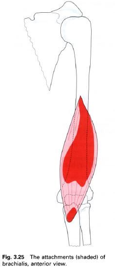
Brachialis lies under cover of biceps brachii in the lower half of the
anterior aspect of the arm. It arises from the distal two-thirds of the anterior
surface of the shaft of the humerus extending outwards onto the
medial and lateral intermuscular septa. The muscle fibers are separated from
the lower part of the lateral intermuscular septum by brachioradialis, with
which it may be partly fused, and extensor carpi radialis longus. The fibres
converge to a thick tendon which forms the floor of the cubital fossa and
attaches to the rough triangular
brachialis impression on the inferior
part of the coronoid process and tuberosity of the ulna. Some deeper fibres of brachialis insert into the capsule of
the elbow joint and serve to pull it away from the moving bones during flexion
and prevent them from becoming trapped.
Nerve
supply
Brachialis is supplied mainly by the musculocutaneous nerve, root value C5
and 6; but also receives a branch from the radial
nerve; again root value C5 and 6, as it runs along the lateral border of
the muscle. However, this latter branch is thought to be almost entirely
sensory.
Action
Brachialis is the main flexor of the elbow
joint.
Functional
activity
Although brachialis flexes the elbow, it is
important in controlling extension produced by gravity. In this situation, the
flexors of the elbow control the movement by an eccentric contraction.
Palpation
When biceps brachii has been identified, brachialis can be felt extending either side
of its belly with the elbow flexed. The tendon can be palpated by applying deep
pressure just above the coronoid process of the ulna.
Brachioradialis
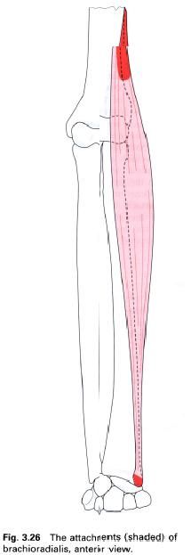
Brachioradialis is a superficial muscle found
on the lateral side of the forearm extending almost as far as the wrist. It
forms the lateral border of the cubital fossa, and is covered in its upper part
by brachialis, with which it may be partly fused. The proximal attachment of
brachioradialis is to the upper
two-thirds of the front of the lateral supracondylar ridge of the humerus and the adjacent part of the lateral intermuscular septum. From here,
the fibres run downwards forming a long, narrow, flat tendon in the middle of
the forearm. The tendon is crossed by those of abductor pollicis longus and
extensor pollicis brevis before it attaches to the lateral surface of the radius just above the styloid process.
Nerve
supply
Brachioradialis is supplied by a branch from
the radial nerve, root value C5 and
6, which enters its medial side above the elbow. The skin over the muscle is
also supplied by roots C5 and 6.
Action
Brachioradialis flexes the elbow joint
particularly when the forearm is midway between pronation and supination. It
also helps to return the forearm to this mid position from the extremes of
either pronation or supination; this can be confirmed by palpation.
Functional
activity
Brachioradialis acts primarily to maintain the
integrity of the elbow joint since its fibres run more or less parallel to the radius. It also works eccentrically as
an extensor of the elbow joint in activities such as hammering.
Palpation
With the elbow flexed to 90° and forearm in a
mid-pronated position, the brachioradialis can be felt along the top of the
forearm when the position is maintained against resistance. Using firm
pressure, the tendon can be palpated proximal to the radial styloid process.
The brachioradialis reflex can be elicited by firmly tapping its tendon just
above the wrist.
Pronator teres
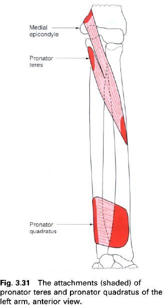
Pronator teres forms the medial border of the
cubital fossa at the elbow, and as such is the most lateral of the superficial
muscles in the flexor compartment of the forearm. It arises by two heads: the humeral head and the ulnar head.
The humeral head arises from the lower part of the medial supracondylar ridge and adjacent intermuscular septum.
It also comes from the common flexor origin on the medial epicondyle and the covering fascia. The ulnar head
arises from the pronator ridge, which
runs downwards from the medial part of the coronoid
process, to join with the humeral head on its deep surface. Between these
two heads passes the median nerve. The muscle fibres pass downwards and
laterally to attach via a flattened tendon into a roughened oval area on the
middle of the lateral surface of the radius.
Nerve
supply
Pronator teres is supplied by the median nerve, root values C6, 7. The
overlying skin is supplied by roots C6 and T1.
Action
Pronator teres pronates the forearm by
producing an anteromedial swing of the
lower end of the radius across the ulna. This movement of the radius carries the hand with it.
Pronator teres is also a weak flexor of the elbow.
Palpation
The muscle can be palpated running along the
medial border of the cubital fossa between the medial epicondyle of the humerus and the middle of the radius. Pronator teres can be most
easily felt, and occasionally seen, when resisting pronation of the forearm.
3. 10. 2012.
Muscles laterally rotating the arm at the shoulder joint
Teres minor
Infraspinatus
Teres minor
When seen from the back, teres minor forms the
upper boundary of both the upper triangular and quadrilateral spaces. It is a
thin muscle which arises by two heads, separated by a groove for the circumflex
scapular artery, from the upper
two-thirds of the lateral border
of the scapula, and the fascia between it and teres major(below) and
infraspinatus(above). The fibres run upwards and laterally forming a narrow
tendon which attaches to the lowest of the facets on the greater tubercle of
the humerus and to the bone
immediately below. The tendon reinforces and blends with the lower posterior
part of the capsule of the shoulder joint.
Nerve
supply
Teres minor is supplied by the axillary nerve, root value C5 and 6. The
skin over the muscle is supplied by roots T1, 2 and 3.
Action
In the anatomical position teres minor is a
lateral rotator, but when the arm is abducted it laterally rotates and adducts.
Palpation
Teres minor can be felt contracting if the
examiner’s fingers are placed half way up the lateral border of the scapula and the arm is then actively
laterally rotated. The tendon is found just below that of infraspinatus as determined
above.

Infraspinatus
Infraspinatus is a thick, triangular muscle
which arises from the medial two-thirds
of the infraspinous fossa of the scapula, tendinous intersections
attached to ridges in this fossa, and the thick fascia covering the muscle. The
fibres converge to a narrow tendon which inserts onto the middle facet on the greater
tubercle of the humerus, and into
the posterior part of the capsule of the shoulder joint. A bursa, which
occasionally communicates with the shoulder joint, separates the muscle from
the neck of the scapula. The upper
part of the muscle lies deep to trapezius,
deltoid and the acromion process;
however, the lower part is superficial.
Nerve
supply
Infraspinatus is supplied by the suprascapular nerve, root value C5 and
6. The skin over the muscle is supplied by the dorsal rami of T1 to T6.
Action
Infraspinatus is a lateral rotator of the arm
at the shoulder joint.
When the arm is laterally rotated, contraction
of infraspinatus can be felt in the medial part of the infraspinous fossa. Its
tendon can be palpated if the greater tubercle is moved from below the acromion
process. To accomplish this the subject lies prone supporting him or herself on
the elbows and forearms. The arm is then laterally rotated some 25° and slightly
adducted. The tendon can now be palpated just below the acromial angle. It is
at this point that soft tissue techniques, such as transverse frictions or
electrical treatments are applied if the tendon becomes inflamed.
Functional
activity
Infraspinatus and teres minor are of importance
during the sequence of movements which occur when the arm is fully abducted.
During the latter part of this movement the humerus
is laterally rotated so that the greater tubercle moves clear of the
coracoacromial arch thereby enabling the remaining part of the humeral head to
come into contact with the glenoid fossa, and full abduction to occur.
Teres minor, infraspinatus, supraspinatus and subscapularis, the musculotendinous “rotator cuff” of extensible
ligaments around the shoulder joint, are all concerned with its stability; the
proximity of their tendons to the joint enhances their effect. During movements
of the head of the humerus on the
glenoid fossa, interplay between these muscles reduces the sliding and shearing
movements which tend to occur. When carrying a weight in the hand these same
four muscles brace the head of the humerus
against the glenoid fossa.
2. 10. 2012.
Muscles medially rotating the arm at the shoulder joint
Subscapularis
Subscapularis
Subscapularis forms the greater part of the
posterior wall of the axilla and as such lies in close proximity to teres major and latissimus dorsi. The anterior surface of the muscle lies on serratus anterior. When seen from the
front it forms the upper boundary of the upper triangular and quadrilateral
spaces.
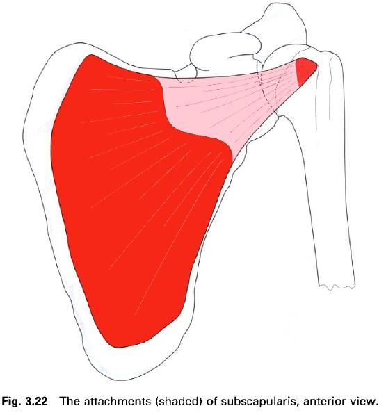
Subscapularis is a multipennate muscle which
arises from the medial two-thirds of
the subscapular fossa and from
tendinous septa, which reinforce the
muscle, attached to bony ridges in the fossa. There is also an attachment to
the fascia covering the muscle. The muscle fibres narrow and form a broad,
thick tendon which attaches to the lesser
tubercle of the humerus, the
capsule of the shoulder joint and the front of the humerus below the tuberosity. A bursa, which communicates directly
with the shoulder joint, separates the tendon from the neck of the scapula.
Nerve
supply
Subscapularis is supplied by the upper and lower subscapular nerves, root
value C5, 6 and 7, from the posterior cord of the brachial plexus.
Action
Subscapularis is a strong medial rotator of the
arm at the shoulder joint; it may also assist in adduction of the arm.
Functional
activity
As part of the “rotator cuff”, subscapularis
plays an important role in maintaining the integrity of the shoulder joint
during movement, by keeping the head of the humerus
within the glenoid fossa. It also resists upward displacement of the humeral
head when deltoid, biceps and the
long head of triceps are active.
Palpation
The muscle belly cannot be palpated as it lies
deep to the scapula. However, careful
deep palpation may allow the tendon to be felt just before its insertion onto
the lesser tuberosity.
1. 10. 2012.
Muscles extending the arm at the shoulder joint
Latissimus dorsi
Teres major
Triceps(long head)
Latissimus dorsi
Latissimus dorsi is a large flat triangular
sheet of muscle running between the trunk, via an extensive attachment, and the
humerus by a narrow tendon. Consequently, it acts on the shoulder joint. The
superior surface of the muscle forms the lower border of the triangle of
auscultation, while its lateral border forms the medial border of the lumbar triangle.

Latissimus dorsi arises from the posterior
layer of the thoracolumbar fascia,
which attaches to the spinous processes
of the lower six thoracic and all of the lumbar and sacral vertebrae,
as well as to the intervening supraspinous
and interspinous ligaments.
That part arising from the lower six thoracic
vertebrae is covered by trapezius. In
addition to this vertebral attachment, latissimus dorsi arises from the
posterior part of the outer lip of
the iliac crest, most laterally by
direct muscular slips. As the muscle fibres sweep upwards and laterally across
the lower part of the thorax, they attach to the outer surfaces of the lower
three or four ribs and via fascia
to the inferior angle of the scapula. From this widespread origin the
fibres converge as they pass to the humerus
and form a thin flattened tendon. The tendon winds around and adheres to
the lower border of teres major, and inserts into the floor of the intertubercular
groove anterior to the tendon of teres major, being separated from it by a
bursa. The effect of twisting the muscle through 180° means that the anterior
surface of the tendon is continuous with the posterior surface of the rest of
the muscle. Consequently, the fibres with the lowest origin on the trunk gain
the highest attachment on the humerus.
Nerve
supply
Latissimus dorsi is supplied by the thoracodorsal nerve, root value C6, 7,
8, which enters the muscle on its deep surface. The skin covering the muscle is
supplied by roots T4 to T12 inclusive, by both ventral and dorsal rami, and L1
to L3 by the dorsal rami.
Action
Latissimus dorsi is a strong extensor of the
flexed arm; however, if the humerus
is fixed relative to the scapula it
retracts the pectoral girdle. It is also a strong adductor and medial rotator
of the humerus at the shoulder joint.
Functional
activity
Functionally, latissimus dorsi is a climbing
muscle, and with the arms fixed above the head it can raise the trunk upwards,
in conjuction with pectoralis major.
Latissimus dorsi has an important function in rowing and during the downstroke
in swimming. Attachment of the muscle to the ribs means that it is active in
violent expiration, and can be felt pressing forcibly inwards during a cough or
sneeze, as it acts to compress the thorax and abdomen.
The attachment to the inferior angle of the scapula allows latissimus dorsi to
assist in holding it against the thorax during movements of the upper limb.
If the humerus
becomes fixed point when standing, as for example when using crutches,
latissimus dorsi is able to pull the trunk forwards relative to the arms;
associated with this is a lifting of the pelvis. In patients with paralysis of
the lower half of the body, the fact that latissimus dorsi attaches to the
pelvis and is still innervated allows it to be used to produce movement of the
pelvis and trunk. Consequently, patients wearing calipers and using crutches
can produce a modified gait by fixing the arms and hitching the hips by the
alternate contraction of each latissimus dorsi.
Palpation
In a lean subject, latissimus dorsi can be made
to stand out relative to the thorax by asking the subject to raise his or her
arm to 90° flexion and to hold it steady against an upwardly directed pressure.
The muscle can be felt contracting if the posterior axillary fold is held
between the finger and thumb while the subject coughs. Adduction of the
abducted arm against resistance also enables latissimus dorsi to be seen and
felt.
Teres major
In the posterior part of the axilla, teres
major forms the lower boundary of both the upper triangular and quadrangular
spaces. It is a thick, chunky muscle, forming, with latissimus dorsi, the
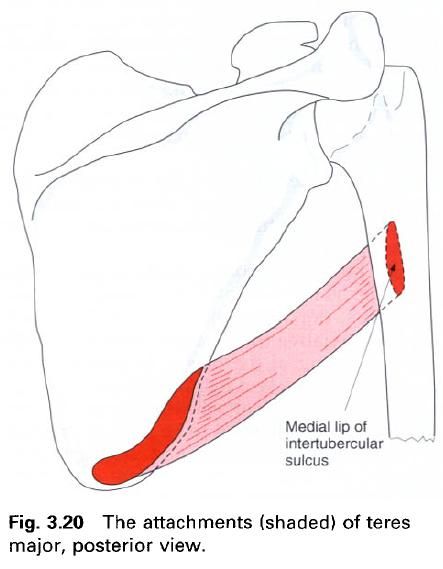
posterior fold of the axilla. It arises from an oval area on the dorsal surface of the scapula near the inferior angle, and from the fascia
between it and adjacent muscles. The muscle fibers, which adhere to those of
latissimus dorsi, run upwards and laterally to form a broad, flat tendon which
attaches along the medial lip of the intertubercular groove. The tendon is
separated from that of latissimus dorsi by a bursa, with the latter muscle
virtually covering the whole of teres major.
Nerve
supply
Teres major is supplied by the lower subscapular nerve, root value C6
and 7.
Action
Teres major adducts and medially rotates the humerus at the shoulder joint. In
addition it can help to extend the flexed arm.
Functional
activity
Teres major, like latissimus dorsi, is a
climbing muscle and works with the latter and pectoralis major to pull the
trunk upwards when the arms are fixed. In conjunction with latissimus dorsi and
pectoralis major, teres major is
important in stabilizing the shoulder joint.
Palpation
Teres major is covered by latissimus dorsi, and
as these two muscles have similar actions, considerable care must be exercised
when it is tested. The inferior angle of the scapula must first be found, the fingers are then moved upwards and
laterally into the posterior wall of the axilla. The subject should abduct the
arm to 90° and then adduct against an upwardly directed resistance. The rounded
contour of teres major should now be palpable. During this same manoeuvre the
flattened tendon of latissimus dorsi, as it twists around teres major, may also
be felt.
Triceps brachii
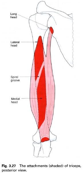
Triceps brachii is situated on the back of the
arm and, as suggested by its name, arises by three heads. Two of the heads
arise from the humerus, being
separated by the spiral groove, and the third comes from the scapula. The three heads are reffered to
as: the long head, lateral head and medial head. The muscle attaches via a
tendon of the ulna.
Long
head. The tendinous long head
comes from the infraglenoid tubercle
of the scapula and the adjacent glenoid labrum, where it blends with the
lower part of the shoulder joint capsule. Named after its length, the long head
is also the most medial of the three, the fibres running downwards superficial
to the medial head before joining the tendon of insertion. As the long head
descends from the infraglenoid tubercle, it passes between teres minor, to
which it is anterior, and teres major, to which it is posterior. In its course
it forms the medial border of the quadrilateral and lower triangular spaces,
and the lateral border of the upper triangular space.
Lateral
head. The fleshy lateral head
arises above and lateral to the spinal groove
on the posterior surface of the humerus between the attachments of teres
minor and deltoid. As the fibres pass
to join with those of the medial head, they cover the spiral groove.
Medial
head. The large, fleshy medial
head lies deep of the other two, and arises from the posterior surface of the humerus,
below and medial to the spiral groove
as far distally as the olecranon fossa. It has an additional attachment to the
posterior aspect of the medial and lateral intermuscular septa.
The three heads of triceps come together to
form a broad, laminated tendon; the superficial part of which covers the
posterior aspect of the lower third of the muscle, while the deeper part arises
from within the substance of the muscle. Such an arrangement creates a larger
surface area for the attachment of the muscle fibres. Both laminae blend to
form a single tendon which attaches to the posterior
part of the proximal surface of
the olecranon of the ulna, and to the deep fascia of the forearm
on either side. Some muscle fibers from the medial head attach to the posterior
part of the capsule of the elbow joint and serve to pull it clear of the moving
bones and prevent it becoming trapped during extension of the joint.
Nerve
supply
All three parts of the muscle are supplied
separately by branches from the radial
nerve. The branch to the lateral head is derived from C6, 7 and 8, while
those to the long and medial heads come from C7 and 8. Of these, the medial
head receives two branches, one of which accompanies the ulnar nerve for a
considerable distance before entering the distal part of the muscle. The other
branch enters more proximally, continuing through the substance of the muscle
to end in, and supply, anconeus. The skin over the muscle is supplied by roots
C5, 7, T1 and T2.
Action
Triceps brachii is the extensor of the elbow
joint. The long head can also adduct the arm and extend it from a flexed
position.
Functional
activity
Once the elbow has been flexed, gravity often
provides the necessary force for extension, with the elbow flexors working
eccentrically to control the movement. Triceps only becomes active in this form
of extension when the speed of the movement becomes important as in executing a
karate chop. Triceps works strongly in pushing and punching activities, and
when performing “press-ups”. In the latter it is working concentrically in the
upward movement, and eccentrically in the downward movement. It works in a
similar manner when using the arms to get out of, or to lower oneself into, a
chair with arms, or when using crutches or parallel bars to relieve body-weight
from the legs during walking. When using a wheelchair, triceps brachii works
strongly to push the wheel round and so propel the chair forwards.
Triceps brachii is also an important extensile
ligament on the under surface of the shoulder joint capsule during abduction of
the arm.
Palpation
The bulk of triceps is easy to see and feel on
the posterior aspect of the arm. All three heads can be felt contracting if the
subject flexes the elbow to 90° with the hand resting on a table, and then
alternately presses downwards and relaxes. The long head can be felt high up on
the back of the arm almost at the axilla; careful palpation enables it to be
traced almost to its insertion on the scapula.
The lateral head can be felt on the upper lateral part of the arm, extending as
far round as the biceps brachii, while the medial head, covered by the other
two heads, can be felt contracting just above the olecranon.
The thick tendon of triceps can be easily
gripped between the thumb and index finger of the examiner’s hand, just above
the olecranon of the ulna. The
triceps reflex is elicited by tapping the tendon just above its insertion, with
the elbow slightly flexed.
Пријавите се на:
Постови (Atom)


 Serbian Dinar Exchange Rate