


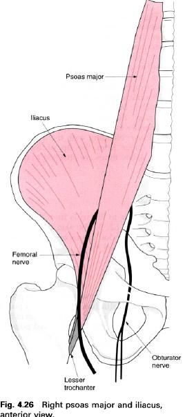
Psoas major
Iliacus
Rectus femoris
Sartorius
Psoas major
Psoas major is a large, thick powerful muscle
situated mainly in the abdominal cavity. Within the substance the lumbar plexus
is found. Psoas major has important relations: at its upper end the diaphragm
and medial arcuate ligament are anterior to psoas, whilst lower down, the
kidney, psoas minor (when present), the renal vessels and ureter are anterior
relations. On the right side, psoas major is overlapped by the inferior vena
cava and the ileum. The ascending and descending colon lie lateral to the right
and left psoas respectively. Medially is the lumbar part of vertebral column,
while directly posterior are the transverse process of the lumbar vertebrae. The
segmental lumbar nerves emerging from the intervertebral foramina are situated
directly behind the muscle and pass forward into its substance.
The upper attachment of psoas major is to the adjacent margins of the bodies of the vertebrae, including the
disc in between. The uppermost attachment is to the lower margin of the body of
the T12, while the lowermost attachment is to the upper margin of the body of
L5. Psoas major also has an attachment to the front and lateral part of
each transverse process, and from tendinous arches covering the
constricted part of the bodies of the lumbar
vertebrae.
The fibres of the muscle pass downwards and
forwards towards the brim of the pelvis. The individual digitations from the
vertebral column join together to form a thick muscle which gradually narrows
as it passes over the pelvic brim under the inguinal ligament. At this point
the tendon changes direction becoming more vertical, it then passes downwards,
backwards and laterally. The muscle is separated from the pubis and the hip joint capsule by a large bursa.
Before passing over the pelvic brim psoas is joined, on its lateral side, by
fibres from iliacus. Iliacus continues to blend with psoas even after the
muscle become tendinous, until it attaches to the tip and posterior aspect of the lesser
trochanter of the femur. Some
fibres of iliacus attach to the femur
on a line running downwards and forwards from the lesser trochanter.
Action
Psoas major is a flexor of the hip joint. It
also, because of its attachment to the lumbar spine, will use the lower
attachment as the fixed point and flex the lumbar spine. There has been much
discussion of the role of this muscle in rotation of the hip joint. It was thought at first that as it attached to the
posterior and medial aspect of the femur,
it must be a lateral rotator of the thigh. However, when the movement of
rotation is around an axis drawn through the head of the femur and the lateral condyle of the tibia, it would clearly be seen that it ought to be a medial
rotator. Electromiographically, however, it shows little activity during either
rotations. The question is still not answered. If psoas major of one side acts
on its own, with reversed origin and insertion, it will produce side flexion of
the lumbar spine to that side.
Nerve
supply
Psoas major is supplied from the anterior rami of L1, 2, 3 and sometimes 4.
The muscle only appears near the surface in the area of the groin and this
small area of skin is supplied by L1.
Functional
activity
The action and functional activity of psoas are
included with those of iliacus as far as flexion of the hip is concerned. However, psoas major does act independently
on the lumbar spine when its lower end is fixed. In sitting up from a lying
position, the muscle on both sides will help to pull the considerable weight of
the trunk up, and this is at a time when the abdominal muscles are working hard
to flex the trunk. It is in fact very important that the abdominal muscles are
brought into action early as this will prevent the lumbar spine being drawn
forwards before the trunk begins to rise. Pulling the head up first will
prevent this unwanted and potentially damaging movement occurring.
Raising both lower limbs at the same time
whilst in the supine position is the cause of much back trouble and,
unfortunately, it has recently become a popular exercise with lay teachers. The
mechanics of this area must be well-understood before exercising is begun as it
is better to prevent back problems rather than try to treat them after they have
occurred.
Each lower limb is approximately 15% of the
body weight, thus when both legs are raised off the floor the hip flexors will
be lifting in the region of 30% of body weight. This initial lift will involve
psoas major, and for about the first 30°, the lumbar spine is pulled upwards
due to the muscle working with reversed origin and insertion. This dragging
forward on the lumbar spine can cause considerable damage to the area,
particularly if some degenerative changes have already occurred. It is believed,
erroneously, that this is a good abdominal exercise and will reduce the
waistline because the abdominal muscles are working hard.
Palpation
It is almost impossible to palpate this muscle
as most of its bulk lies within the abdominal cavity. It appears near the
surface in the groin, but it is still quite difficult to feel as it is covered
by other structures.
Iliacus
Iliacus is a large fleshy triangular muscle
situated mainly in the pelvis. Its upper attachment is the larger coming mainly
from the upper and posterior two-thirds of the iliac fossa with some fibres coming from the ala of the sacrum and anterior
sacroiliac ligament. Its fibres pass downwards, forwards and medially
blending with the lateral side of psoas major. This blending of the muscles
continues over the pelvic brim where they change direction to pass downwards,
backwards and slightly laterally to insert into the lesser trochanter of the femur, blending with the insertion of the
psoas major from the tip of the lesser trochanter. A few fibres are attached to
the hip joint capsule.
Nerve
supply
The nerve supply to iliacus is from the femoral nerve, root value L2, 3. The
skin covering the area, where the tendon passes over the brim of the pelvis, is
supplied by L1.
Action
Its effect on the hip is similar to that of psoas major. If its upper attachment is
the fixed point it will pull the thigh forwards as in flexion of the hip. If the lower attachment is the
fixed point it will draw the pelvis forwards, thus tilting it forwards; being
again flexion of the hip but this
time with the trunk doing the moving.
Functional
activity
This muscle will be used with psoas major in
all activities of pulling the lower limb up in front of the trunk as in drawing
the lower limb forward in walking, running and jumping. It will also, of
course, help to draw the trunk forward from a lying supine position to a
sitting position. There is the same controversy over rotation of the femur as for the psoas major.
Palpation
It is almost impossible to palpate( similar to
psoas major).
Rectus femoris
Rectus femoris is seen to stand out on the
front of the thigh, being a spindleshaped bipennate muscle. Its upper
attachment is by two heads which are continuous with each other, one to the anterior inferior iliac spine (this is
the straight head) and the other to a rough area immediately above the acetabulum (this is the reflected head).
It is thought that the straight head is a human acquisition associated with the
evolution of an upright posture. From this continuous origin, a single tendon
descends from which arises the fleshy belly of the muscle.

About two thirds of the way down the thigh, the
muscle begins to narrow to a thick tendon attaching to the upper border of the patella.
From here, some fibres pass around the patella
helping to form the ligamentum patellae. The deep surface of the muscle is
tendinous and smooth, allowing free movement over a similar surface on vastus
intermedius, thus permitting its independent action on the hip joint.
Sartorius
Sartorius is the most superficial muscle in the
anterior compartment of the thigh. Its lower part is mainly on the medial side
anterior to gracilis. It is a long
strap-like muscle having flattened tendons at each end. It is renowned as the
longest muscle in the body, getting its name from its action, which is to
produce most of the actions needed in the lower limb, to produce cross-legged
sitting, the position that tailors used to use when making clothes.
It upper attachment is to the anterior superior
iliac spine and the area just below. From here the muscle passes medially and
inferiorly to attach to a vertical line on the medial side of the shaft of the tibia, in front of both semitendinosus and gracilis, partly blending with the latter. A few fibres from the
lower tendon go to the medial collateral ligament of the knee joint and to the
fascia of the leg. A bursa separates sartorius from gracilis at its lower end. The medial border of the upper third
forms the lateral boundary of the femoral triangle whilst the middle third
forms the roof of the adductor canal.

Nerve
supply
Sartorius is supplied by the femoral nerve, root value L2, 3. The
area of skin covering this muscle is also supplied by L2, 3.
Action
Sartorius will produce many of the movements
which are combined to produce cross-legged sitting, that is flexion of the hip and knee, lateral rotation and
abduction of the thigh, and medial rotation of the tibia on the femur.
These actions can be summarized by saying that it places the heel on the medial
side of the opposite knee.
Functional
activity
Going into the cross-legged or the tailor
sitting position is a functional activity; however, sartorius will help to
produce any activity which involves flexion of the knee and hip together, combined with lateral
rotation of the thigh as in drawing up the lower limbs when using the breast
stroke in swimming.
Palpation
Sartorius is most easily palpated at its
proximal end just below the anterior superior iliac spine. Here its strap-like
shape can be easily palpated, particularly when the leg, with the knee slightly
flexed, is raised some 15cm from the floor when lying supine.


0 коментара:
Постави коментар