


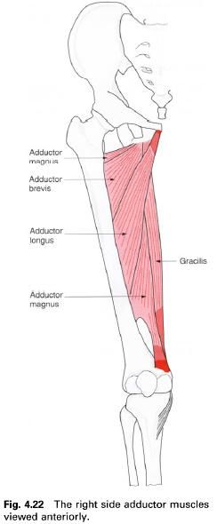
Adductor magnus
Adductor longus
Adductor brevis
Gracilis
Pectineus
These muscles are situated on the medial side
of the hip joint running down the
medial side of the thigh.
Adductor magnus
Adductor magnus is the largest and most
posterior of the group, with adductor brevis and adductor longus in front, and semimembranosus and semitendinosus
behind. The muscles is really composed of two parts, an adductor part and a hamstring part, forming a large
triangular sheet of muscle with a thickened medial margin.
Its upper attachment is from the femoral surface of the ischiopubic ramus running down to the
lateral part of the inferior surface
of the ischial tuberosity. The part
of the muscle which attaches anteriorly to the ischiopubic ramus represents a
sheet of muscle which twists before attaching to the femur while the posterior fibres of adductor magnus, from the
ischial tuberosity, pass vertically downwards as a thickened cord of muscle
fibres.
The ischiopubic fibres fan out and are seen as
a large triangular muscular sheet. The most anterior of these fibres pass
laterally and slightly backwards to attach to the upper part of the linea
aspera continuing upwards as far as the greater
trochanter medial to the attachment of gluteus maximus. These upper fibres may be fused with quadratus femoris. Fibres
from the posterior part of the ischiopubic ramus attach to the whole length of
the linea aspera and medial supracondylar
ridge. This attachment to the femur
is not continuous as there are small fibrous arches close to the bone which
allow vessels and nerves to pass from the adductor to the posterior compartment
of the thigh. The posterior ischial fibres pass downwards and attach mainly to
the adductor tubercle situated on top
of the medial condyle of the femur at
the lower end of the medial supracondylar ridge. Some of these fibres continue
downwards to fuse with the medial collateral ligament of the knee.
Nerve
supply
Because of its two parts, adductor magnus has a
dual nerve supply. The adductor part from the ischiopubic ramus is supplied by
the posterior division of the obturator
nerve (L2, 3), while the hamstring
part from the ischial tuberosity is supplied by the tibial division of the sciatic nerve (L4), the skin covering the
inner side of the thigh being mainly from L3.
Action
Working as a whole, the muscle is an adductor
of the hip joint; although the
posterior portion will aid in extension of the hip. Some people believe that this muscle, together with the
adductor longus, medially rotates the hip
joint, although it was believed in the past that they also acted as lateral
rotators. Whether the muscle acts acted a medial or lateral rotator will depend
on the position of the thigh, and the line of action of the muscle with respect
to the mechanical axis of the femur.
All the adductor muscles are important in preventing lateral overbalancing
during the support phase of walking.
It is worth noting that the medial collateral
ligament of the knee joint appears to be a downwards continuation of the
adductor magnus tendon and as such this muscle may very well at some time have
crossed the knee joint and therefore have been a flexor of the knee in a
similar fashion to gracilis.
Palpation
Adductor magnus is a deep muscle and is therefore
difficult to palpate, nevertheless, if the fingers are pushed in just above the
medial condyle of the femur, the
adductor tubercle can be identified. If the inside of the same foot is now pressed against a stationary
obstacle, one can feel the vertical part of the muscle contracting. The muscle
can be traced about one-third of the way up the thigh until it becomes hidden
by other muscles.
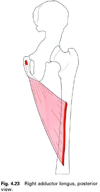
Adductor longus
Adductor longus is a long, slender, triangular
muscle situated on the medial aspect of the thigh, overlying the middle part of
adductor magnus. Its upper, narrower attachment comes from a small roughened
area just below the medial end of the obturator
crest on the anterior aspect of the body of the pubis.
Its fibres pass downwards and laterally
spreading out as they go to attach to the middle two-quarters of the linea aspera, anterior to adductor
magnus below and adductor brevis above and posterior to vastus medialis.
Nerve
supply
Adductor longus is supplied by the anterior division of the obturator nerve,
root value L2, 3, 4. The skin covering the area of adductor longus is supplied
by L3.
Action
Adductor longus is an adductor of the thigh,
but as a rotator of the thigh there is some doubt. Adductor longus can also
flex the extended thigh, and extend the flexed thigh.
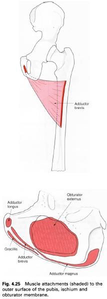
Adductor brevis
Adductor brevis is again a triangular muscle
situated on the medial aspect of the thigh.
Its upper attachment is from the lateral part of the front of the body and inferior ramus of the pubis.
Its fibres pass downwards, laterally and backwards to attach to the upper half
of the linea aspera anterior to
adductor magnus. Its upper part is posterior to pectineus and its lower part is
posterior to adductor longus.
Nerve
supply
Adductor brevis is supplied by the anterior division of the obturator nerve,
root value L2, 3, 4. The skin covering the area of adductor brevis is supplied
by L2.
Action
Adductor brevis is an adductor of the thigh.
Palpation
If the fingers are placed high up on the inside
of the thigh and the lower limb is adducted against resistance a mass of muscle
can be palpated running down towards the thigh. These are the adductors;
however it is difficult to distinguish between the different muscle masses.
The adductors
Gracilis is the most medial muscle of the
adductor group. It is an adductor of the thigh as well as flexor of the knee.
Functional
activity
Although it is quite clear that these muscles
are adductors of the thigh, they appear to work most strongly when the hip joint is in the neutral position,
that is the anatomical position. They certainly work strongly, synergically,
when the knee and hip are being
flexed and extended when weight-bearing. There is still some confusion over
whether these muscles are involved in either medial or lateral rotation of the
thigh. They work strongly during walking, as they pull the supporting leg into
adduction, thereby moving the line of gravity over the supporting foot. They also contribute to the
delicate balancing of the pelvis on the hip
joint. The adductors, as a group, are used very strongly when some object is
being held between the knees in the sitting position, for example, when sitting
on a horse, particularly when the horse is moving.
Gracilis
Gracilis, as its name implies, is a long, thin
muscle, situated on the medial side of the thigh. It is the most superifical of
the adductor group. Its upper attachment is to the front of the body of the pubis and its inferior ramus,
just encroaching on to the ramus of
the ischium. The muscle, as it
descends between semimembranosus
posteriorly and sartorius anteriorly, develops a fusiform-shaped belly at about
its middle. It becomes tendinous above the knee and crosses the joint before
expanding to attach to a short vertical line on the upper part of the medial
surface of the shaft of the tibia.
This attachment is above that of semitendinosus
and behind and blending with that of sartorius. Bursae separate the tendon of
gracilis from those of sartorius and semitendinosus.

Nerve
supply
Gracilis is supplied by the anterior division of the obturator nerve,
root value L2, 3. The skin covering this area is innervated by roots L2, 3; the
upper part by the obturator nerve and the lower part by the femoral nerve.
Action
Although this muscle is situated with the adductor
group of muscles, its action of adduction on the thigh is not so important as
its action on the knee. It is mainly a flexor of the knee, but with the knee in
a semiflexed position it will aid medial rotation of the leg on the thigh.
Functional
activity
As a flexor of the knee this muscle will help the hamstrings in simple flexion
activities, such as the beginning of the swing phase in walking when the knee
needs to be flexed. It will also help when strong flexion is required, as when
pulling the body forward on the sliding seat of a rowing boat. In horse riding,
gracilis is used in all its actions. When the rider is gripping the horse,
gracilis will help the adductor muscles, whilst at the same time helping to
control the flexed knee.
Palpation
In the sitting position with the medial aspect
of the foot against a solid object,
such as the leg of a table, or when the toes are inwardly rotated, the tendon
can be felt on the posteriomedial aspect of the knee joint, being the upper of
the two obvious tendons. If traced upwards, the belly of the muscle can be
palpated and traced to its attachment on the front of the pubic body.
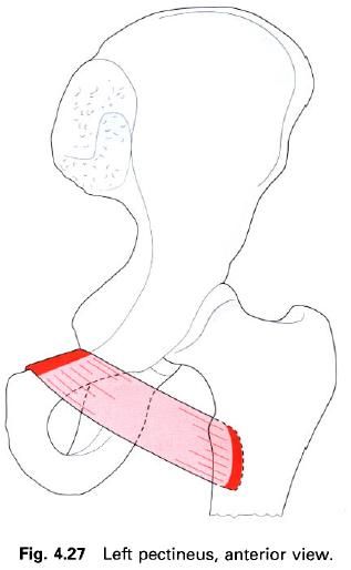
Pectineus
Pectineus is a quadrilateral muscle situated at
the upper and medial part of the thigh, deep in the groin. It appears to be
made up of two layers, superficial and deep, and these are generally supplied
by different nerves.
Its upper attachment is to the superior ramus (pectin) of the pubis, the iliopubic eminence and the pubic
tubercle. It also attaches to the fascia
which covers it. The fibres pass downwards, backwards and laterally between
psoas major and adductor longus to attach to a line which runs from the lesser trochanter of femur to the top of
the linea aspera, anterior to the
upper part of adductor brevis. This is often called the pectineal line.
Nerve
supply
Pectineus is supplied by the femoral nerve from its L2, 3 nerve roots
and occasionally from the obturator or
the accessory obturator nerve by its L3 root. The skin covering this area
of the groin is supplied from the root of L1.
Action
Pectineus flexed and adducts the hip joint. Some authorities also believe
the muscle to be a medial rotator of the hip.
Functional
activity
It is easy to see how this muscle acts as a
flexor and adductor of the hip by
looking at the direction of its fibres, which pass downwards, backwards and
laterally. Contraction of the muscle will therefore draw the thigh inwards and
forwards. Most authorities dismiss the rotation element as there is certainly
disagreement, although some feel that there must be more to the rotation than
has yet been deduced.
There is no doubt that the insertion of
pectineus is lateral and in front of the mechanical axis of the femur (the line around which rotation
would occur in the standing position). Thus the muscle in this case would
produce medial rotation. However, when the foot
is off the ground, as in the carrying-through phase in walking, the axis would
still pass through the hip joint, but
now would vary considerably according to the position of the thigh and also
that of the pelvis. In fact it would depend very much on the swing of the lower
limb.
So far, the functional activity of this muscle
has only been considered in the upright, standing or walking position. Much of
the time is spent in the sitting position with the hip joint flexed at a right angle. The relationship between the
origin and insertion of this muscle is now completely reversed. To make the
situation even clearer, the thigh can be raised off the seat until it is at an
angle of 45° to the horizontal – as if the legs were going to be crossed. The
muscle fibres now pass forwards and upwards, passing well behind the axis of
the rotating thigh. The action of the muscle in this position will now be
adduction as before, but also extension and lateral rotation; in fact a
movement very similar to that of crossing the legs except that the thigh is
being pulled down on to the opposite thigh. This movement is comparable with
the initial stages of rising from a very low chair, or from the squat position,
especially if the movement is being carried out at some speed and under load.
If the argument is taken one stage further, the
muscle is obviously a flexor and adductor in the upright position with perhaps
medial rotation.
The muscle is an extensor and lateral rotator
in the fully flexed position, but still performing adduction. Therefore, as in
the case of many muscles in the body, pectineus can perform different actions
according to its starting position and the relative position of its origin and
insertion. It is not surprising, therefore, that it is supplied by nerves from
both the flexor and adductor compartments of the thigh.
Remembering the dual nerve supply, the dual
action, the closeness of the muscle to the hip
joint and its important relations, it is surprising that pectineus only merits
a few lines in most anatomy texts. It must have played a vital role in
locomotion with a flexed hip, either
in climbing or when all four limbs were on the ground. Has its role diminished
that much or are we overlooking the true action and worth of pectineus?


0 коментара:
Постави коментар