


|
Quadriceps femoris
|
|
|
Vastus lateralis
|
|
|
Vastus medialis
|
|
|
Vastus intermedius
|
Quadriceps femoris is the large muscle bulk on
the anterior surface of the thigh. As its name implies, it is composed of four
main parts. One part, rectus femoris, has its origin above the hip joint, while the other three parts take origin from the
shaft of the femur. All four join
together around the patella to form a
thick strong tendon called the ligamentum patellae, which inserts into the
tibial tuberosity.
Vastus lateralis
Vastus lateralis is situated on the
anterolateral aspect of the thigh, lateral to rectus femoris. It has an
extensive linear attachment from the upper
lateral part of the intertrochanteric
line, the lower border of the greater trochanter, the lateral side of the gluteal tuberosity and the upper
half of the lateral lip of the linea aspera. It also attaches to the
fascia lata and lateral intermuscular septum. From this origin the muscle
fibres run downwards and forwards with those at the top passing almost
vertically downwards.
The muscle bulk is mainly situated in the upper
half of the lateral side of the thigh, from which a broad tendon arises which
narrows down as it approaches the lateral side of the patella. This tendon inserts into the tendon of rectus femoris and the base and lateral border of the patella. Some fibres pass to the front of the
lateral condyle of the tibia blending
with the iliotibial tract helping to form the expansion which finally attaches
to the line running towards the tibial tuberosity. To a large extent this part
of its attachment replaces the knee joint capsule in this region.
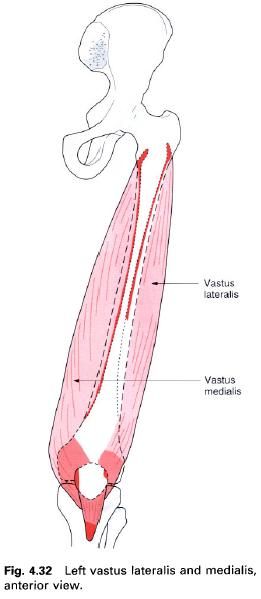
Vastus medialis
Vastus medialis is situated on the anteromedial
aspect of the thigh, medial to rectus femoris, with most of its bulk showing at
the lower third just above the patella.
It has an extensive linear origin from a line beginning at the lower medial end of the intertrochanteric line, running downwards
around the medial aspect of the upper end of the shaft on the spiral line, the medial lip of the linea aspera, continuing on to the upper two-thirds of the medial supracondylar line, the medial
intermuscular septum and the tendon of adductor magnus.
Its upper fibres pass mainly downwards, whilst
its lower fibres tend to pass almost horizontally forwards. These two sets of
fibres which make up vastus medialis are considered by some to be anatomically
and functionally distinct, with the oblique fibres being called vastus medialis
obliquus. The muscle attaches to the tendon
of rectus femoris, the medial border
of the patella, and the front of the medial condyle of the tibia. The
expansions which pass across the knee joint to attach to the tibia replace the joint capsule in this region and become fused
with the deep fascia. This attachment also runs to the tibial tuberosity.
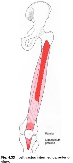
Vastus intermedius
Vastus intermedius is the deepest part of
quadriceps femoris lying between vastus lateralis and medialis, and deep to
rectus femoris.
It arises by fleshy fibres from the upper
two-thirds of the anterior and lateral surfaces of the femur, its fibres pass downwards to form a broad tendon on its
more superficial aspect. This attaches to the deep surface of the tendons of
rectus femoris and the other vastus muscles, and to the base of the patella. In
the middle of the thigh, vastus intermedius is difficult to separate from
vastus lateralis, while lower down it is impossible to separate from vastus
medialis.
Articularis
genus. Some of the deep fibres
of vastus intermedius, arising from a small area on the inferior third of the anterior surface of the femur, pass downwards to attach to the upper part of the suprapatellar
bursa of the knee joint which lies deep to vastus intermedius. These fibres are the articularis genus. Its
main function is to prevent the synovial membrane becoming trapped and
interfering with the normal movements of the knee joint.
Ligamentum patellae
All four of quadriceps tendons contribute to the
formation of the ligamentum patellae. It runs from the apex of the patella to the upper part of the tibial
tuberosity acting as the tendon of insertion of the quadriceps femoris
muscle. The patella is really a sesamoid bone in the tendon of rectus femoris
and vastus intermedius helping to relay the pull of the quadriceps over the
front of the femur.
Nerve
supply
Quadriceps femoris, including articularis
genus, is supplied by the femoral nerve,
root value L2, 3, 4. The skin covering the quadriceps is supplied by L2, 3.
Quadriceps femoris
Action
Although rectus femoris is part of the
quadriceps femoris group, by crossing anterior to the hip joint it also flexes the thigh.
Quadriceps femoris is the main extensor of the
knee joint. Rectus femoris crosses in front of the hip joint and therefore is also a flexor of that joint. Each muscle
appears to have its particular role in extension of the knee and often comes in
at different ranges of the movement. For example, vastus medialis is more
obviously active in the final stage of extension and is believed to resist the
tendency of the patella to move
laterally caused by the angulation of the femur. Specific exercises designed to strengthen the oblique fibres of
vastus medialis are advocated by some practitioners in order to affect tracking
of the patella, resisting dislocation
and possibly helping reduce anterior knee pain in certain circumstances.
Rectus femoris works particularly strongly in
straight leg rising or in the combined movement of flexion of the hip and extension of the knee.
Functional
activity
The quadriceps are used strongly in stepping
activities, for example stair climbing and squats. Rectus femoris will perform
its function particularly in the swing phase of walking when the lower limb is
being carried forward and the knee is being extended. Vastus medialis, in the
final stages of extension of the knee, will help in the locking mechanism of
the joint when the femur is allowed
to rotate medially.
Surprisingly, in the standing position very little
or no action is recorded in the quadriceps as in this position the knees are in
the closepacked position. It is at these times that if the knees are knocked
from behind, forward collapse will almost certainly occur. However, when
standing on a moving vehicle the quadriceps will be active. When standing on
one leg, all the muscles around the knee will work statically to provide
stability at the joint.
The quadriceps is a powerful and an important
muscle. It must work strongly throughout its full range. It will lose strength
and bulk rapidly if there is any injury to it or to the knee joint. It may take
months to regain power, but only days to lose it.
Palpation
When sitting on a chair and straightening the
knee joint, particularly against resistance, the separate parts of quadriceps
femoris, except vastus intermedius, can easily be palpated: medialis on the
lower medial aspect, lateralis in the upper half of the lateral side and rectus femoris running down the centre. Stand with the knees semiflexed, place your
hands on the front of each thigh; the three parts of the muscle (as above) can
be readily palpated.
In straight leg raising, the muscle should be
able to extend the knee into a few degrees of hyperextension, this being the
extra range required for the knee to be able to lock. Patients not being able
to do this will often complain of their knee giving way during walking.


1 коментара:
Thanks for sharing the post.Jhansi orthopedic hospital provides best fracture treatment in jhansi.
Постави коментар