


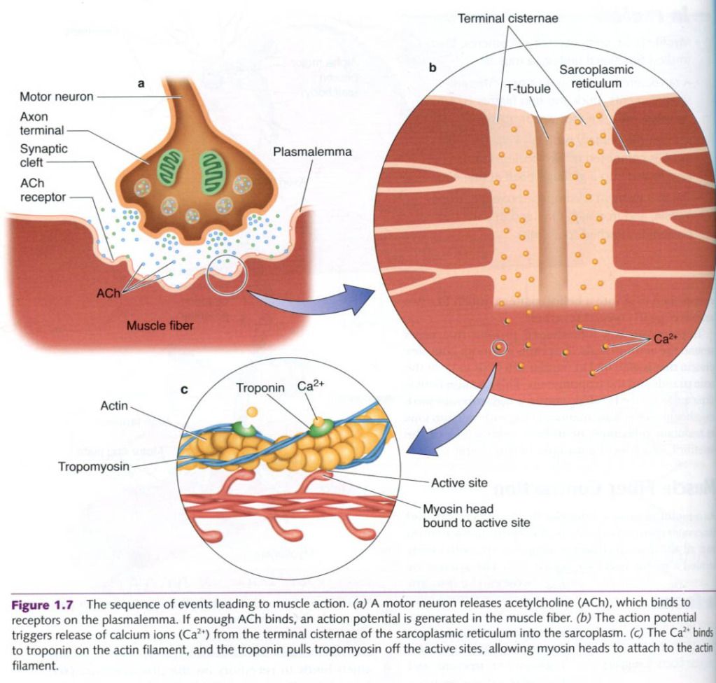
Action potential
The events that trigger a muscle fiber to
contract are complex. The process, that is described on the second picture, is
inititated by an electric signal, or action
potential, from the brain or spinal cord to an alpha-motor neuron. The
action potential arrives at the alpha-motor neuron’s dendrites, specialized
receptors on the neuron’s cell body. From here, the action potential travels
down the axon to the axon terminals, which are located very close to the
plasmalemma. When the action potential arrives, these nerve endings secrete a
neurotransmitter substance called acetylcholine(Ach), which binds to receptors
on the plasmalemma(second picture, part a). If enough Ach binds to the
receptors, the action potential will be transmitted the full length of the
muscle fiber as ion gates open in the muscle cell membrane and allow sodium to
enter. This process is reffered to as depolarization. An action potential mus
be generated in the muscle cell before the muscle cell can act.
Role
of calcium in the muscle fiber
In addition to depolarizing the fiber membrane,
the action potential travels over the fiber’s network of tubules(T-tubules) to
the interior of the cell. The arrival of an electrical charge causes the
adjacent SR to release large quantities of stored calcium ions(Ca2+)
into the sarcoplasm.
In the resting state, tropomyosin molecules
cover the myosin-binding sites on the actin molecules, preventing the binding
of the myosin heads. Once calcium ions are released from the SR, they bind to
the troponin on the actin molecules. Troponin, with its strong affinity for
calcium ions, is believed to then initiate the contraction process by moving
the tropomyosin molecules off the myosin-binding sites on the actin molecules.
This is shown in the picture 2,part c. Because tropomyosin normally cover the
myosin-binding sites, it blocks the attraction between the myosin cross-bridges and actin molecules. However, once the
tropomyosin has been lifted off the binding sites by troponin and calcium, the
myosin heads can attach to the binding sites on the actin molecules.
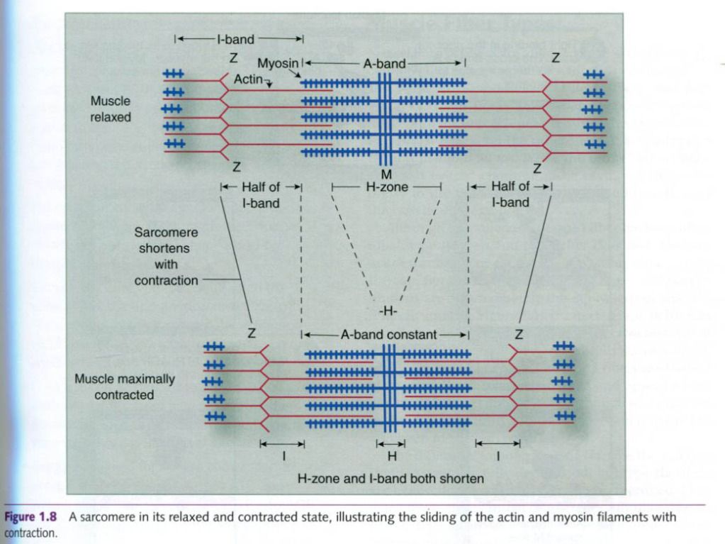
Sliding filament theory: How muscle creates movement
When muscle contracts, muscle fibers shorten.
How do they shorten? The explanation for this phenomenon termed the sliding filament theory. When the
myosin cross-bridges are activated, they bind with actin, resulting in a
conformational change in the cross-bridge, which causes the myosin head to tilt
and drag the thin filament toward the center of the sarcomere. This tilting of
the head is reffered to as the power
stroke. The pulling of the thin filament past the thick filament
shortens the sarcomere and generates force. When the fibers are not
contracting, the myosin head remains in contact with the actin molecule, but
the molecular bonding at the site is weakened or blocked by tropomyosin.
Immediately after the myosin head tilts, it
breaks away from the active site, rotates back to its original position, and
attaches to a new active site farther along the actin filament. Repeated
attachments and power strokes cause the filaments to slide past one another,
giving rise to the term sliding filament
theory. This process continues until the ends of the myosin filaments reach
the Z-disks, or until the Ca2+ is pumped back into the sarcoplasmic
reticulum. During this sliding(contraction), the thin filaments move toward the
center of the sarcomere and protrude into the H-zone, ultimately overlapping.
When this occurs, the H-zone is no longer available.
Energy
for muscle contraction
Muscle contraction is an active process
requiring energy. In addition to the binding site for actin, a myosin head
contains a binding site for adenosine
triphosphate(ATP). The myosin molecule must bind with ATP for muscle
contraction to occur because ATP supplies the needed energy.
The enzyme adenosine
triphosphatase(ATPase), which is located on the myosin head, splits the ATP
to yield adenosine diphosphate(ADP), inorganic phosphate(Pi), and energy. The
energy released from this breakdown of ATP is used to power the tilting the
myosin head. Thus, ATP is the chemical source of energy for muscle contraction.
End
of muscle contraction
Muscle contraction continues as long as calcium
is available in the sarcoplasm. At the end of a muscle contraction, calcium is
pumped back into the SR, where it is stored until a new action potential
arrives at the muscle fiber membrane. Calcium is returned to the SR by an
active calcium-pumping system. This is another energy-demanding process that
also relies on ATP. Thus,energy is required for both the contraction and
relaxation phases.
When the calcium is pumped back into the SR,
troponin and tropomyosin return to the resting conformation. This blocks the
linking of the myosin cross-bridges and actin molecules and stops the use of
ATP. As a result, the thick
and thin filaments return to the their original relaxed state.
The molecular events of a contractile cycle –
changes in the myosin head during various phases of the powerstroke:
1) Tight
binding in the rigor state. The cross-bridge is at a 45 degrees angle relative
to the filaments.
2) ATP
binds to its binding site on the myosin. Myosin then dissociates from actin.
3) The
ATPase activity of myosin hydrolyzes the ATP. ADP and Pi remain bound to
myosin.
4) The
myosin head swings over and binds weakly to a new actin molecule. The
cross-bridge is now at 90 degrees relative to the filaments.
5) Release
of Pi initiates the power stroke. The myosin head rotates on its hinge, pushing
the actin filament past it.
6) At
the end of the power stroke, the myosin head releases ADP and resumes the
tightly bound rigor state.
“Physiology of sport and exercise”, fourth edition; Jack H. Wilmore, David L. Costill, W. Larry Kenney
“Physiology of sport and exercise”, fourth edition; Jack H. Wilmore, David L. Costill, W. Larry Kenney


0 коментара:
Постави коментар